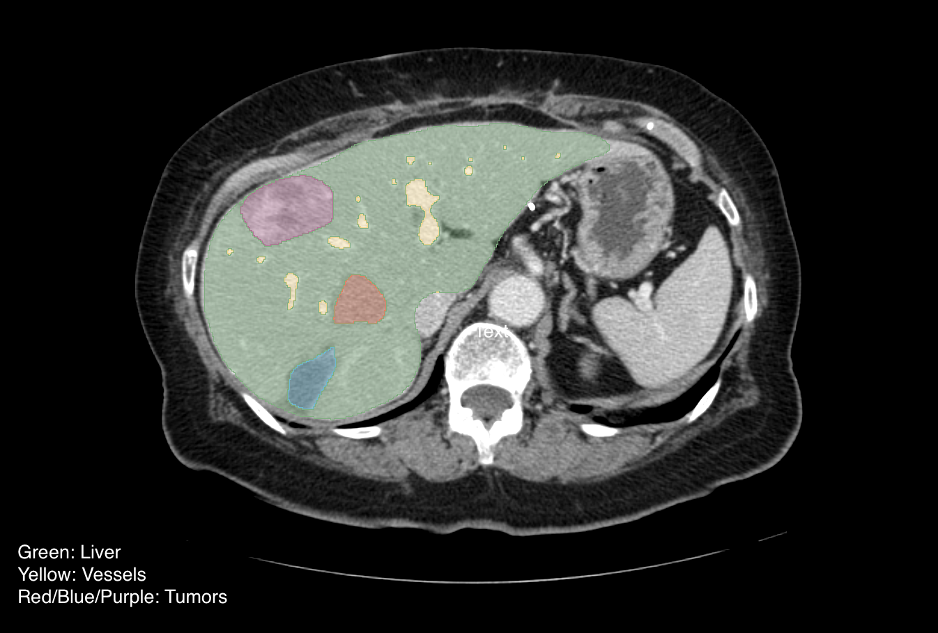
PHANTOM-FDA

By
DOI: 10.7937/k9/TCIA.2015.orbjkmux | Image Collection

As part of a more general effort to probe the interrelated factors impacting the accuracy and precision of lung nodule size estimation, we have been conducting phantom CT studies with an anthropomorphic thoracic phantom containing a vasculature insert on which synthetic nodules were inserted or attached.

The utilization of synthetic nodules with known truth regarding size and location allows for bias and variance...

Read More

PELVIC-REFERENCE-DATA

By
DOI: 10.7937/TCIA.2019.WOSKQ5OO | Image Collection

Purpose: Expert selected landmark points on clinical image pairs provide a basis for rigid registration validation. Using combinatorial rigid registration optimization (CORRO) we provide a statistically characterized reference data set for image registration of the pelvis by estimating the optimal ground truth.

Methods: Landmark points for each CT/CBCT image pair for 58 pelvic...

Read More

PEDIATRIC-CT-SEG

By
DOI: 10.7937/TCIA.X0H0-1706 | Image Collection

This dataset was collected by a collaboration of researchers from Children’s Wisconsin, Marquette University, Varian Medical Systems, Medical College of Wisconsin, and Stanford University as part of a project funded by the National Institute of Biomedical Imaging and Bioengineering (U01EB023822) to develop tools for rapid, patient-specific CT organ dose estimation. The collection consists of CT images in DICOM format...

Read More

PANCREAS-CT

By
DOI: 10.7937/K9/TCIA.2016.tNB1kqBU | Image Collection

The National Institutes of Health Clinical Center performed 82 abdominal contrast enhanced 3D CT scans (~70 seconds after intravenous contrast injection in portal-venous) from 53 male and 27 female subjects.  Seventeen of the subjects are healthy kidney donors scanned prior to nephrectomy.  The remaining 65 patients were selected by a radiologist from patients who neither had major abdominal pathologies nor pancreatic...

Read More

OSTEOSARCOMA-TUMOR-ASSESSMENT

By
DOI: 10.7937/tcia.2019.bvhjhdas | Image Collection

Osteosarcoma is the most common type of bone cancer that occurs in adolescents in the age of 10 to 14 years. The dataset is composed of Hematoxylin and eosin (H&E) stained osteosarcoma histology images. The data was collected by a team of clinical scientists at University of Texas Southwestern Medical Center, Dallas. Archival samples for 50 patients treated at Children’ s Medical Center, Dallas, between 1995...

Read More

NSCLC-RADIOMICS-INTEROBSERVER1

By
DOI: 10.7937/tcia.2019.cwvlpd26 | Image Collection

This collection contains clinical data and computed tomography (CT) from 22 non-small cell lung cancer (NSCLC) radiotherapy patients. For 21 of these patients with pre-treatment CT scans, repeated blinded manual delineations by five different radiation oncologists of the 3D volume of the gross tumor volume on CT and clinical outcome data are available. The above was repeated with the same set of five radiation oncologists,...

Read More

NSCLC-RADIOGENOMICS

By
DOI: 10.7937/K9/TCIA.2017.7hs46erv | Image Collection

Medical image biomarkers of cancer promise improvements in patient care through advances in precision medicine. Compared to genomic biomarkers, image biomarkers provide the advantages of being a non-invasive procedure, and characterizing a heterogeneous tumor in its entirety, as opposed to limited tissue available for biopsy. We developed a unique radiogenomic dataset from a Non-Small Cell Lung Cancer (NSCLC)...

Read More

NRG-1308

By
DOI: 10.7937/K9/TCIA.2016.6fC8z46u | Image Collection

This data collection consists of images acquired in preparation for NRG-1308, a phase 3 randomized trial to compare overall survival after photon versus proton chemoradiation therapy for inoperable stage II-IIIB NSCLC. The images include IMRT photon and proton scans. A detailed summary of the full trial from which these patients were included...

Read More