
CC-RADIOMICS-PHANTOM-3

By
DOI: 10.7937/tcia.2019.j71i4fah | Image Collection

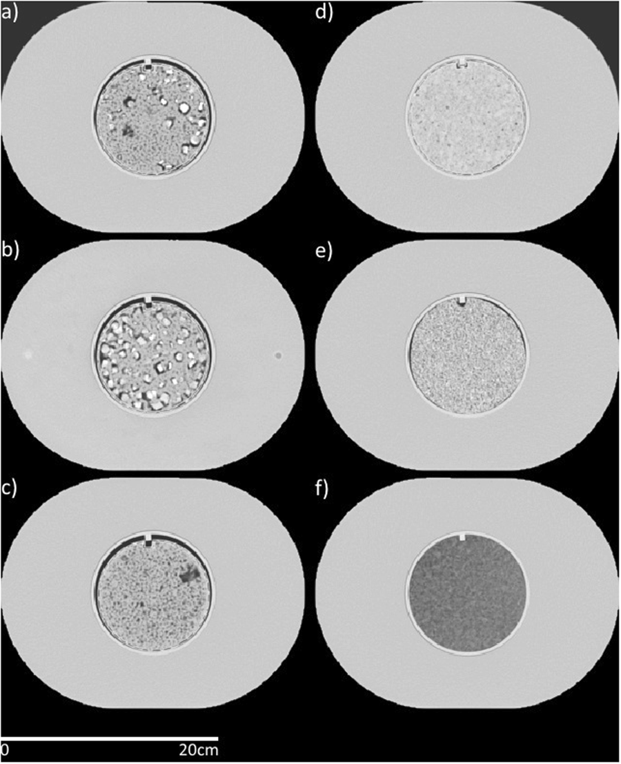
There currently is a dearth of phantom scans on large samples. This data collection contains one physical phantom, imaged across three protocols, on 100 scanners. This provides population data that can be used to quantify inter-scanner variability. This data can be used to determine how robust specific radiomics or other quantitative imaging signatures are.

Protocol

Computed tomography scans were acquired...

Read More

CT-IMAGES-IN-COVID-19

By
DOI: 10.7937/TCIA.2020.GQRY-NC81 | Image Collection

These retrospective NIfTI image datasets consists of unenhanced chest CTs: 

  • First dataset - from 632 patients with COVID-19 infections at initial point of care, and
  • Second dataset - a second set of 121 CTs from 29 patients with COVID-19 infections with serial / sequential CTs.

The initial images for both datasets were acquired at the point of care in an outbreak setting from patients...

Read More

CC-RADIOMICS-PHANTOM-2

By
DOI: 10.7937/TCIA.2019.4l24tz5g | Image Collection

This collection consists of 251 CT scans of Credence Cartridge Radiomic (CCR) phantom. This texture phantom was developed to investigate the feature robustness in the emerging field of radiomics. This phantom dataset was acquired on 4-8 CT scanners using a set of imaging parameters (e.g., reconstruction Field of View, Slice thickness, reconstruction kernels, mAs, and Pitch). A controlled scanning approach was employed...

Read More

CC-RADIOMICS-PHANTOM

By
DOI: 10.7937/K9/TCIA.2017.zuzrml5b | Image Collection

This collection consists of 17 CT scans of the Credence Cartridge Radiomics (CCR) phantom, which was designed for use in studies of texture feature robustness. The scans were acquired at four medical centers using each center’s chest protocol and were taken using GE (7 scans), Philips (5 scans), Siemens (2 scans), and Toshiba (3 scans) scanners. The CCR phantom has 10 cartridges, each with a unique texture, Fig 1....

Read More

C-NMC-2019

By
DOI: 10.7937/tcia.2019.dc64i46r | Image Collection

Acute lymphoblastic leukemia (ALL) constitutes approximately 25% of the pediatric cancers. In general, the task of identifying immature leukemic blasts from normal cells under the microscope is challenging because morphologically the images of the two cells appear similar.

Challenge is split into 3 separate phases:

  • Train set composition:

    Total subjects:...

Read More

COVID-19-AR

By
DOI: 10.7937/tcia.2020.py71-5978 | Image Collection

Radiology imaging  is playing an increasingly vital role in the diagnosis of COVID-19 patients and determining therapeutic options, patient care management and new research directions. Publicly available imaging data is essential to drive new research by permitting the creation of large multi-site cohorts for machine learning based analyses.  All too frequently rural populations are underrepresented in such public...

Read More