CT COLONOGRAPHY | ACRIN 6664
DOI: 10.7937/K9/TCIA.2015.NWTESAY1 | Data Citation Required | Image Collection
| Location | Species | Subjects | Data Types | Cancer Types | Size | Status | Updated | |
|---|---|---|---|---|---|---|---|---|
| Colon | Human | 825 | CT | Colon Cancer | Image Analyses | Public, Complete | 2013/11/15 |
Summary
The National CT Colonography Trial (ACRIN 6664) collection contains 825 cases of CT colonography imaging with accompanying spreadsheets that provide polyp descriptions and their location within the colon segments. Additional information about the trial is available in the Study Protocol and Case Report Forms. Main Objective: To clinically validate widespread use of computerized tomographic colonography (CTC) in a screening population for the detection of colorectal neoplasia. Participants: Male and female outpatients, aged 50 years or older, scheduled for screening colonoscopy, who have not had a colonoscopy in the past five years. Study Design Summary: The study addresses aspects of central importance to the clinical application of CTC in several interrelated but independent parts that will be conducted in parallel. In Part I, the clinical performance of the CTC examination will be prospectively compared in a blinded fashion to colonoscopy. In Part II, optimization of the CT technique will be performed in view of new technological advances in CT technology. In Part III, lesion detection will be optimized by studying the morphologic features of critical lesion types and in the development of a database for computer-assisted diagnosis. In Part IV, patient preferences and cost-effectiveness implications of observed performance outcomes will be evaluated using a predictive model.
Data Access
Version 1: Updated 2013/11/15
| Title | Data Type | Format | Access Points | Subjects | License | |||
|---|---|---|---|---|---|---|---|---|
| Radiology Images | CT | DICOM | Download requires NBIA Data Retriever |
825 | 836 | 3,451 | 941,771 | CC BY 3.0 |
| DICOM Metadata Digest | CSV | CC BY 3.0 | ||||||
| Polyp Descriptions - Large 10mm | XLS | CC BY 3.0 | ||||||
| Polyp Descriptions - 6 to 9mm | XLS | CC BY 3.0 | ||||||
| Polyp Descriptions - No polyp found | XLS | CC BY 3.0 |
Additional Resources for this Dataset
The NCI Cancer Research Data Commons (CRDC) provides access to additional data and a cloud-based data science infrastructure that connects data sets with analytics tools to allow users to share, integrate, analyze, and visualize cancer research data.
- Imaging Data Commons (IDC) (Imaging Data)
Citations & Data Usage Policy
Data Citation Required: Users must abide by the TCIA Data Usage Policy and Restrictions. Attribution must include the following citation, including the Digital Object Identifier:
Data Citation |
|
|
Smith K, Clark K, Bennett W, Nolan T, Kirby J, Wolfsberger M, Moulton J, Vendt B, Freymann J. (2015). Data From CT COLONOGRAPHY. The Cancer Imaging Archive. https://doi.org/10.7937/K9/TCIA.2015.NWTESAY1 |
Detailed Description
There are presently 825 cases in this collection with XLS sheets that provide polyp descriptions and their location within the colon segments. To link the XLS polyp tables with the DICOM image studies in TCIA you should understand that some cases in the TCIA are identified by long numbers with the last 4 digits after the last decimal point (e.g.: NCIA study number “1.3.6.1.4.1.9328.50.4.0040” referred to as case “40”). In addition there are a fewer number of additional positive cases that begin their identification number with ‘CTC’ (e.g.: CTC-5401799343)
Three related XLS spreadsheets are in this release.
- TCIA CTC large 10 mm polyps.xls – Contains the case numbers for 35 cases (out of the 825 total TCIA cases) where at least one 10mm or larger size polyp was found. Individual cases may have several (up to 20) polyps of different sizes listed on a particular XLS row as “LESION 1.x, 2.x,3.x etc. – see “feature key” below).
- TCIA CTC 6 to 9 mm polyps.xls – Contains 69 cases with smaller size polyps.
- TCIA CTC no polyp found.xls – Contains 243 cases that were recorded as free of polyps by both CTC and optical techniques.
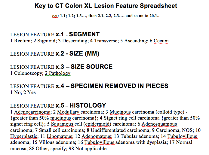
Thus in this CT Colonography collection you will be able to download the prone and supine DICOM images from OC same-day validated 243 negative cases, 69 cases with 6 to 9 mm polyps, and 35 cases which have at least one > 10 mm polyp and their histological type. Below is the key for deciphering the features in the spreadsheet.
WARNING: NCI cannot assure archive users of error-free validity of the XL polyp location data since NCI did not itself perform the clinical study or its analysis.
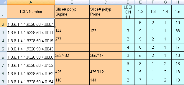
You will note that two XLS files with positive findings have multiple columns descriptors of individual polyp lesions listed as in the table below. The meaning of the colored columns labeled “LESION 1.1…1.2…1.3…1.4, etc” is explained in the attached key-code “.tiff” file entitled “Polyp description key table.tiff”). Some CT scan slice numbers where the polyps were found are provided, but unfortunately the table may not have complete slice number information – you’ll just have to do the best you can with the data NCI was given.
A note about the values in DICOM header (0010,1010) Patient Age: Within the CT COLONOGRAPHY collection, there are some ages that were anonymized to 105Y and 110Y which does not follow the standard TCIA de-identification procedure regarding ages 89 and older. TCIA policy is to bin any age over 89 into a common age group “090Y”. Although these ages are not binned to 090Y, please be aware that they are not actual ages of patients.
Related Publications
Publication Citation |
|
|
Johnson, C. D., Chen, M.-H., Toledano, A. Y., Heiken, J. P., Dachman, A., Kuo, M. D., … Limburg, P. J. (2008, September 18). Accuracy of CT Colonography for Detection of Large Adenomas and Cancers. New England Journal of Medicine. New England Journal of Medicine (NEJM/MMS). https://doi.org/10.1056/nejmoa0800996 |
Research Community Publications
A full list of publications that reference this dataset can be found on Dimensions.ai (free log-in required)
TCIA maintained a legacy list of publications through 2023 found below and on the TCIA Publications Page.
- Shakir, Hina; Deng, Yiming; Rasheed, Haroon; Khan, Tariq Mairaj Rasool. Radiomics based likelihood functions for cancer diagnosis. Sci Rep 2019 link
- Gayathri, Devi K; Radhakrishnan, R; Rajamani, Kumar. Segmentation of colon and removal of opacified fluid for virtual colonoscopy. Pattern Analysis and Applications 2017 link
- Lin, Anthony Y; Du, Peng; Dinning, Philip G; Arkwright, John W; Kamp, Jozef P; Cheng, Leo K; Bissett, Ian P; O’Grady, Gregory. High-resolution anatomic correlation of cyclic motor patterns in the human colon: Evidence of a rectosigmoid brake. American Journal of Physiology-Gastrointestinal and Liver Physiology 2017 link
- Manjunath, KN; Siddalingaswamy, PC; Prabhu, GK. Measurement of smaller colon polyp in CT colonography images using morphological image processing. International journal of computer assisted radiology and surgery 2017 link
- Alazmani, A; Hood, A; Jayne, D; Neville, A; Culmer, P. Quantitative assessment of colorectal morphology: Implications for robotic colonoscopy Medical engineering & physics 2016 link
- Manjunath, KN; Siddalingaswamy, PC; Gopalakrishna Prabhu, K. An improved method of colon segmentation in computed tomography colonography images using domain knowledge Journal of Medical Imaging and Health Informatics 2016 link
- Yahya-Zoubir, Bahia; Hamami, Latifa; Saadaoui, Llies; Ouared, Rafik. Automatic 3D Mesh-Based Centerline Extraction from a Tubular Geometry Form. Information Technology And Control 2016 link
- Gayathri Devi, K; Radhakrishnan, R. Automatic Segmentation of Colon in 3D CT Images and Removal of Opacified Fluid Using Cascade Feed Forward Neural Network. Computational and Mathematical Methods in Medicine 2015 link
- Manjunath, KN; Siddalingaswamy, PC; Prabhu, GK. Automatic Electronic Cleansing in Computed Tomography Colonography Images using Domain Knowledge. Asian Pacific Journal of Cancer Prevention 2015 link
- Namías, R; D’Amato, JP; Del Fresno, M; Vénere, M. Automatic rectum limit detection by anatomical markers correlation Computerized Medical Imaging and Graphics 2014 link
- Boone, Darren J; Halligan, Steve; Roth, Holger R; Hampshire, Tom E; Helbren, Emma; Slabaugh, Greg G; McQuillan, Justine; McClelland, Jamie R; Hu, Mingxing; Punwani, Shonit. CT Colonography: External Clinical Validation of an Algorithm for Computer-assisted Prone and Supine Registration. Radiology 2013 link
- Roth, Holger R; Boone, Darren J; Halligan, Steve; Hampshire, Thomas E; McClelland, Jamie R; Hu, Mingxing; Punwani, Shonit; Taylor, Stuart; Hawkes, David J. External clinical validation of prone and supine CT colonography registration. 2012 link
The Collection authors suggest the below will give context to this dataset: