CT Images in COVID-19 | CT Images in COVID-19
DOI: 10.7937/TCIA.2020.GQRY-NC81 | Data Citation Required | Image Collection
| Location | Species | Subjects | Data Types | Cancer Types | Size | Status | Updated |
|---|---|---|---|---|---|---|---|
| Lung | Human | 661 | CT | COVID-19 (non-cancer) | Public, Complete | 2021/05/25 |
Summary
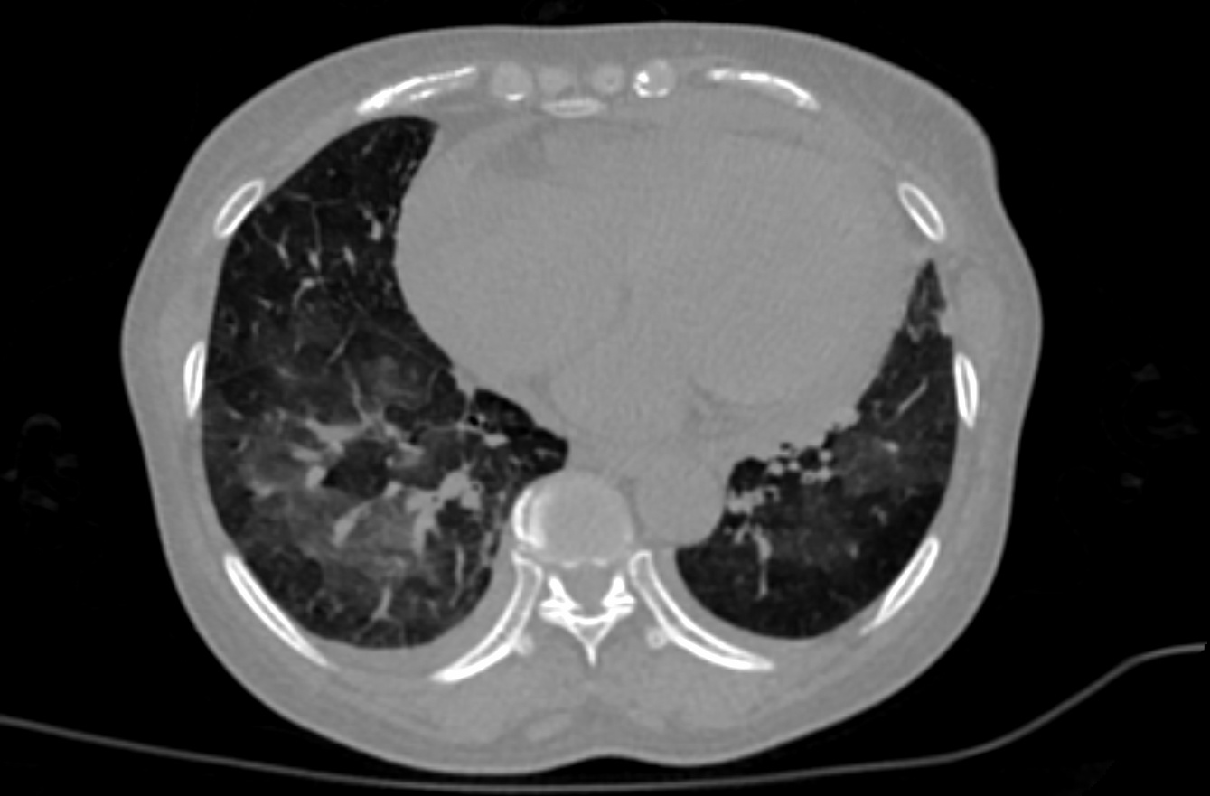
These retrospective NIfTI image datasets consists of unenhanced chest CTs: The initial images for both datasets were acquired at the point of care in an outbreak setting from patients with Reverse Transcription Polymerase Chain Reaction (RT-PCR) confirmation for the presence of SARS-CoV-2. A multidisciplinary team trained several models using portions of the first dataset, along with additional CTs and manually annotated images from other sources. A classification model derived in part from the first dataset is described in a Nature Communications manuscript at: https://doi.org/10.1038/s41467-020-17971-2. The NVIDIA-related frameworks and models specific to this publication are available at no cost as part of the NVIDIA Clara Train SDK at https://ngc.nvidia.com/catalog/containers/nvidia:clara:ai-covid-19. This includes both inference-based pipelines for evaluation, as well as model weights for further training or fine tuning in outside institutions. The second data set of 121 serial / sequential CTs in 29 patients is reported in a Scientific Reports manuscript at https://doi.org/10.1038/s41598-021-85694-5. 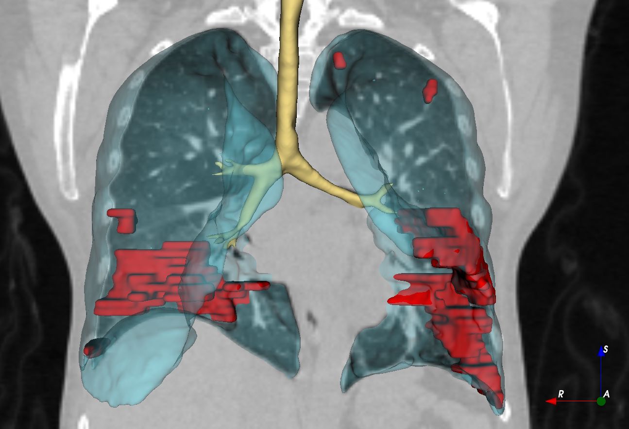 Patients presented to a health care setting with a combination of symptoms, exposure to an infected patient, or travel history to an outbreak region. All patients had a positive RT-PCR for SARS-CoV-2 from a sample obtained within 1 day of the initial CT. CT exams were performed without intravenous contrast and with a soft tissue reconstruction algorithm. The DICOM images were subsequently converted into NIfTI format. The second dataset also had other follow up CTs, in addition to the initial point of care CT.
Data Access
Version 2: Updated 2021/05/25
Added second dataset, 29 patients/121 CT images.
| Title | Data Type | Format | Access Points | Subjects | License | |||
|---|---|---|---|---|---|---|---|---|
| Images First dataset | CT | NIFTI | Download requires IBM-Aspera-Connect plugin |
632 | 0 | 650 | CC BY 4.0 | |
| Images Second dataset | CT | NIFTI | Download requires IBM-Aspera-Connect plugin |
29 | 121 | CC BY 4.0 |
Citations & Data Usage Policy
Data Citation Required: Users must abide by the TCIA Data Usage Policy and Restrictions. Attribution must include the following citation, including the Digital Object Identifier:
Data Citation |
|
|
An, P., Xu, S., Harmon, S. A., Turkbey, E. B., Sanford, T. H., Amalou, A., Kassin, M., Varble, N., Blain, M., Anderson, V., Patella, F., Carrafiello, G., Turkbey, B. T., & Wood, B. J. (2020). CT Images in COVID-19 [Data set]. The Cancer Imaging Archive. https://doi.org/10.7937/TCIA.2020.GQRY-NC81 |
Acknowledgement |
|
|
The Multi-national NIH Consortium for CT AI in COVID-19. |
Acknowledgements
The Imaging AI in COVID team would like to acknowledge the following individuals who supported this multi-disciplinary multi-national team effort:
- All frontline workers and Peng An, Sheng Xu, Evrim B. Turkbey, Stephanie A. Harmon, Thomas H. Sanford, Amel Amalou, Michael Kassin, Nicole Varble, Maxime Blain, Dilara Long, Dima Hammoud, Ashkan Malayeri, Elizabeth Jones, Holger Roth, Ziyue Xu, Dong Yang, Andriy Myronenko, Victoria Anderson, Mona Flores, Francesca Patella, Maurizio Cariati, Kaku Tamura, Hirofumi Obinata, Hitoshi Mori, Ulas Bagci, Daguang Xu, Hayet Amalou, Robert Suh, Gianpaolo Carrafiello, Baris Turkbey, Bradford J. Wood.
- Thanks for leadership support to: John Gallin, Steve Holland, Cliff Lane, Bruce Tromberg, Tom Misteli, Bill Dahut.
- Supported by the NIH Center for Interventional Oncology and the NIH Intramural Targeted Anti-COVID-19 (ITAC) Program.
TCIA COVID-19 Datasets
Additional datasets and information about TCIA efforts to support COVID-19 research can be found here.
Related Publications
Publication Citation |
|
|
Link to publication below contains AI model that was only partly derived from this data, and also from other data not present here on TCIA: Harmon, S. A., Sanford, T. H., Xu, S., Turkbey, E. B., Roth, H., Xu, Z., Yang, D., Myronenko, A., Anderson, V., Amalou, A., Blain, M., Kassin, M., Long, D., Varble, N., Walker, S. M., Bagci, U., Ierardi, A. M., Stellato, E., Plensich, G. G., Franceschelli, G., Girlando, C., Irmici, G., Labella, D., Hammoud, D., Malayeri, A., Jones, E., Summers, R. M., Choyke, P.L., Xu, D., Flores, M., Tamura, K., Obinata, H., Mori, H., Patella, F., Cariati, M., Carrafiello, G., An, P., Wood, B. J., & Turkbey, B. (2020). Artificial intelligence for the detection of COVID-19 pneumonia on chest CT using multinational datasets. Nature Communications, 11(1). https://doi.org/10.1038/s41467-020-17971-2
|
Research Community Publications
TCIA maintains a list of publications which leverage our data. If you have a manuscript you’d like to add please contact the TCIA Helpdesk.