This collection is a set of head and neck cancer patients' multiple positron emission tomography/computed tomography (PET/CT) 18F-FDG scans–before and after therapy–with follow up scans where clinically indicated. The data was provided to help facilitate research activities of the National Cancer Institute's (NCI's) Quantitative Imaging Network (QIN). This collection was supported by Grants: U24 CA180918 (http://qiicr.org)...
This collection contains “double baseline” multi-parametric MRI images collected on patients with newly diagnosed glioblastoma. The value of this collection is to provide clinical image data to establish the test-retest characteristics of parameters calculated from DW-MRI, DCE-MRI, and DSC-MRI such as ADC, Ktrans and rCBV. Data were provided by Dr. Elizabeth Gerstner and Dr. Kalpathy-Cramer (MGH) as part of their...
This data is from a multi-site, multi-parametric quantitative MRI study of adult (18+ years old) females diagnosed with invasive breast cancer. Subjects all had a lesion size >1cm in longest dimension and were undergoing neoadjuvant therapy. Participants were scanned prior to any therapy and then 2-3 times after the initiation of neoadjuvant therapy, depending upon their treatment regimen. All data sets were...
The QIBA CT-1C phantom collection was designed and shared to assist in Characterizing Variability, sans Biology. This data set was contributed by RSNA's Quantitative Imaging Biomarker Alliance activity, Volumetric CT Group 1C. Multiple image sets of the same phantoms...
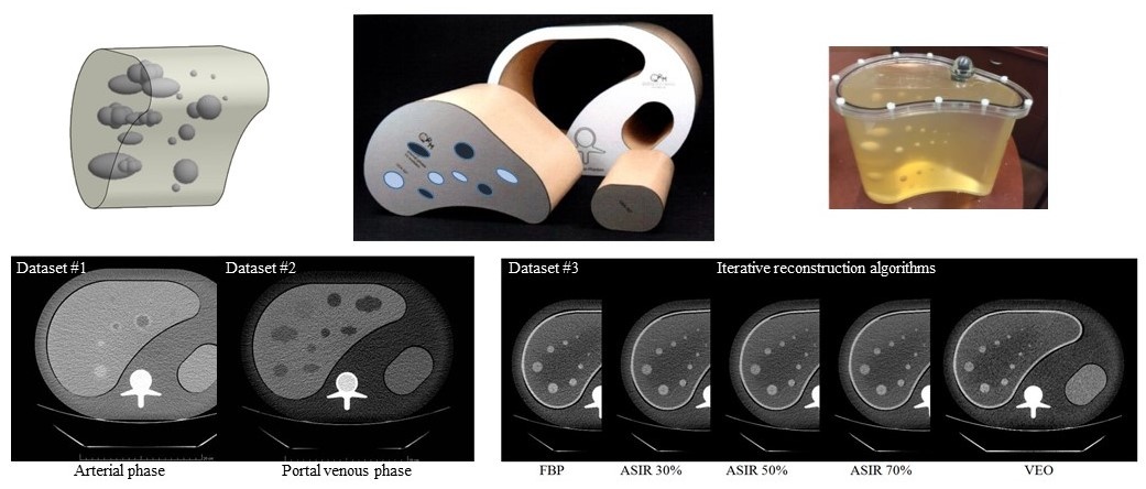
This database contains a collection of three sets of CT scan images acquired from an anthropomorphic abdominal phantom with removable liver inserts. The anthropomorphic phantoms were designed by a group of scientists from FDA and Columbia University Medical Center and custom manufactured by QRM (Moehrendorf, Germany).
Data sets #1 (AP phantom) & #2 (PVP phantom): Two liver inserts, each containing 19 embedded synthetic...
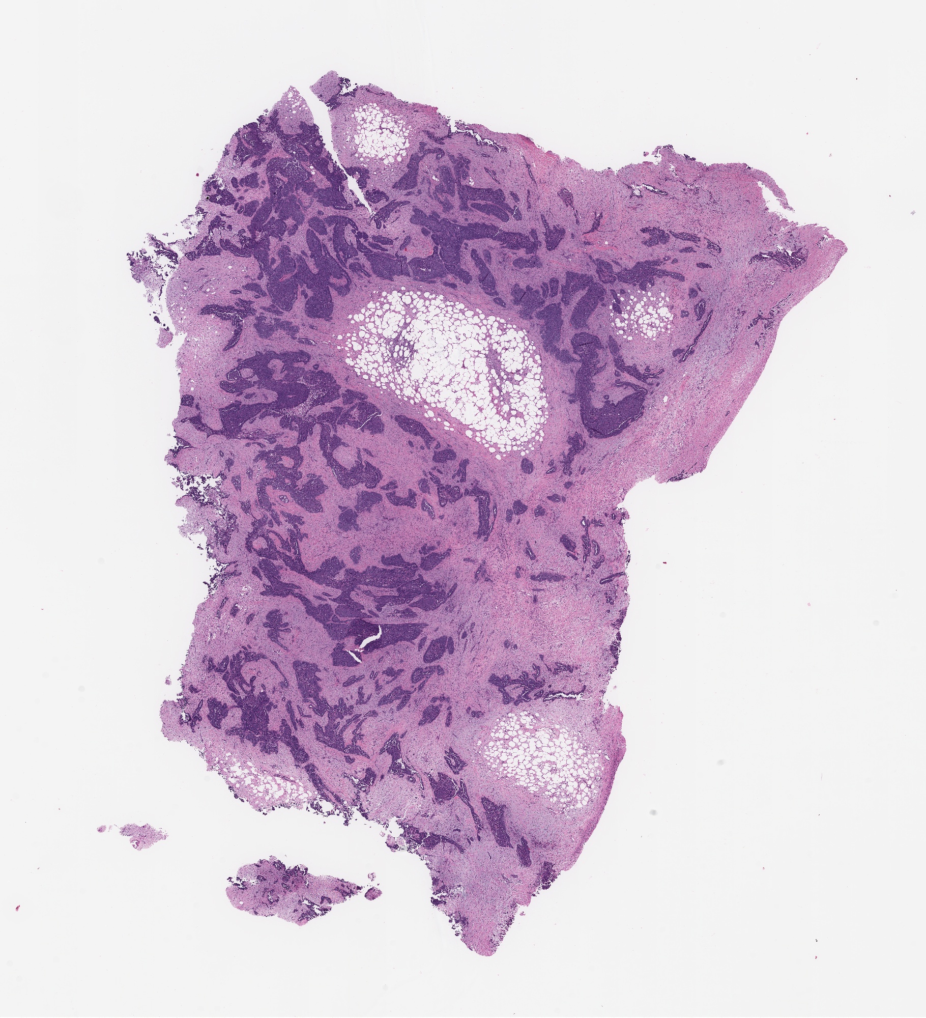
In our study, we have generated proteomic and genomic (RNA sequencing and whole genome sequencing) profiles from high grade serous ovarian cancer (HGSOC) tumor biopsies. All biospecimens are formalin-fixed, parrafin-embedded (FFPE) tissues and annotated for patient sensitivity to platinum chemotherapy (refractory or sensitive). For all 174 tumors that were analyzed, we have H&E-stained and imaged the first and...
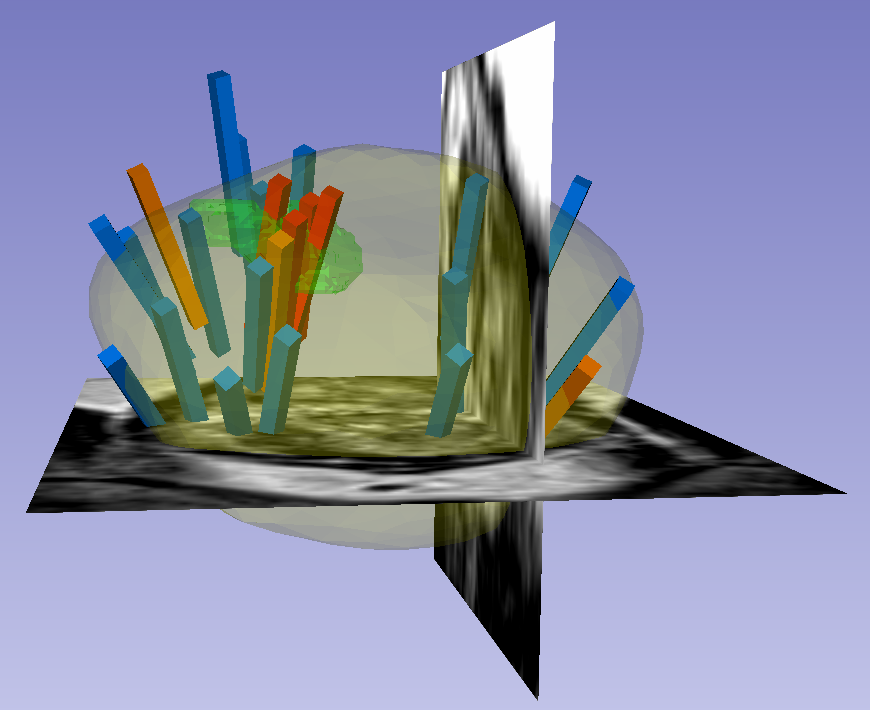
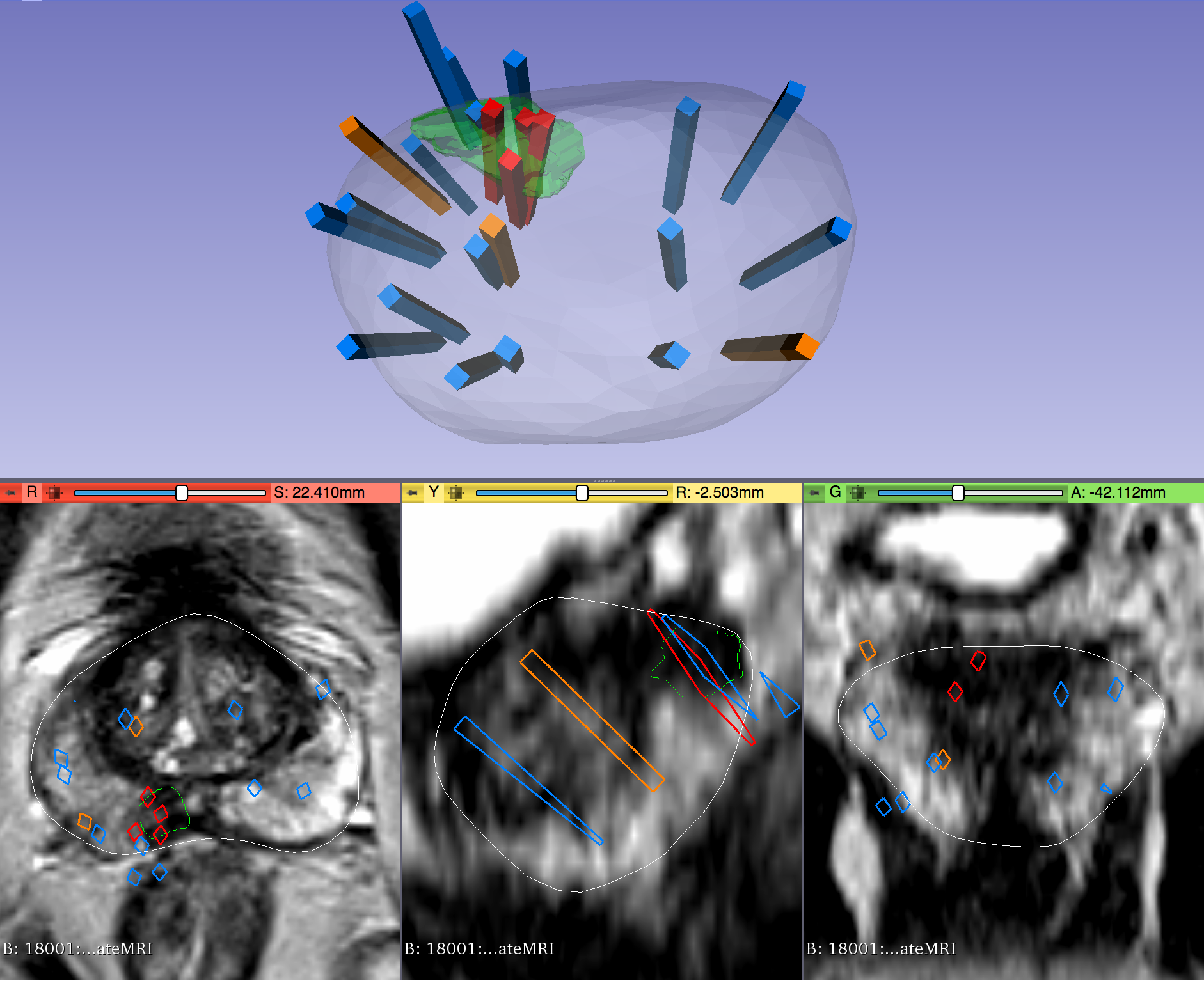
This dataset was derived from tracked biopsy sessions using the Artemis biopsy system, many of which included image fusion with MRI targets. Patients received...
This collection of prostate Magnetic Resonance Images (MRIs) was obtained with an endorectal and phased array surface coil at 3T (Philips Achieva). Each patient had biopsy confirmation of cancer and underwent a robotic-assisted radical prostatectomy. A mold was generated from each MRI, and the prostatectomy specimen was first placed in the mold, then cut in the same plane as the MRI. The data was generated at the National...
Prostate cancer T1- and T2-weighted magnetic resonance images (MRIs) were acquired on a 1.5 T Philips Achieva by combined surface and endorectal coil, including dynamic contrast-enhanced images obtained prior to, during and after I.V. administration of 0.1 mmol/kg body weight of Gadolinium-DTPA (pentetic acid). Corresponding clinical metadata (XLS format) and 3D segmentation files (NRRD format) are offered as a supplement...