QIBA-CT-Liver-Phantom | QIBA Anthropomorphic Abdominal Phantom CT Scans
DOI: 10.7937/TCIA.RMV0-9Y95 | Data Citation Required | Image Collection
| Location | Species | Subjects | Data Types | Cancer Types | Size | Status | Updated | |
|---|---|---|---|---|---|---|---|---|
| Liver Phantom | Human | 3 | CT | Phantom | Image Analyses | Public, Complete | 2021/04/27 |
Summary
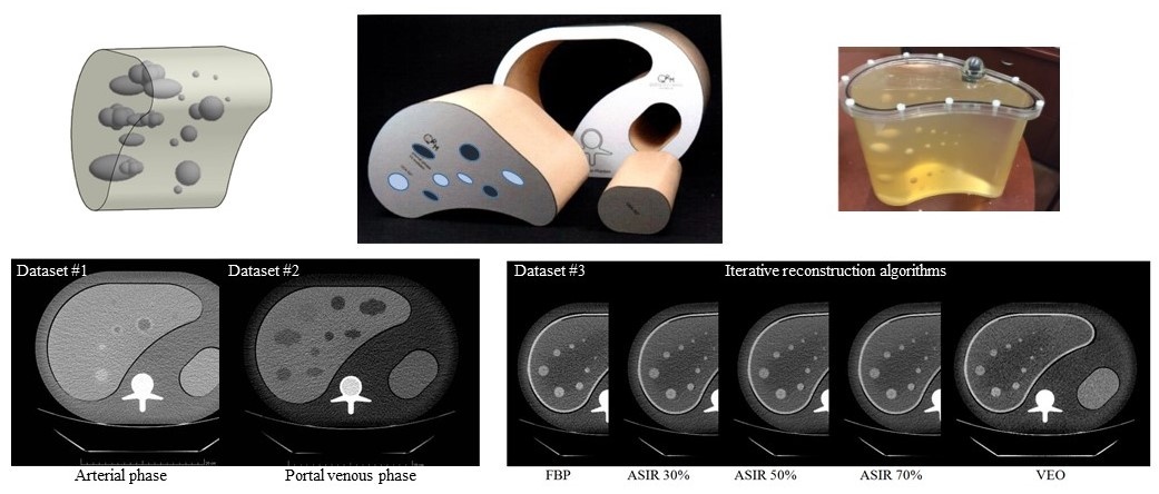
This database contains a collection of three sets of CT scan images acquired from an anthropomorphic abdominal phantom with removable liver inserts. The anthropomorphic phantoms were designed by a group of scientists from FDA and Columbia University Medical Center and custom manufactured by QRM (Moehrendorf, Germany). Data sets #1 (AP phantom) & #2 (PVP phantom): Two liver inserts, each containing 19 embedded synthetic lesions with known volumes of varying diameter (6–40 mm), shape, contrast (10–65 HU), and density (homogenous and mixed) were designed to have liver parenchyma and lesion CT values simulating arterial phase (AP phantom) and portal venous phase (PVP phantom) imaging, respectively. The two phantoms were scanned using two 64-slice multi-detector helical CT scanners (GE 750HD and Siemens Biograph mCT) across a wide range of abdominal imaging protocols, including three effective mAs (50, 100, 250), two pitches (GE/Siemens: 1.375 and 0.983/1.35 and 1.0), four slice thicknesses (GE/Siemens: 0.625, 1.25, 2.5, and 5 mm/0.6, 1.5, 3, and 5 mm), and two convolution kernels (GE/Siemens: Standard and Soft/B20f and B30f). Two repeated scans were performed for each protocol and for each phantom. Dataset #3 (IR phantom): One non-uniform liver insert was filled with an in-house made gelatin-based background, and varied concentrations of gelatin, salt, and water were used to mimic normal liver tissue and focal fat at the arterial phase. Ten spherical lesions of five sizes (20, 14, 10, 7, and 5 mm in diameter) and two radiodensities (95 and 110 HU) were placed on the border between the fatty and normal parenchyma. The phantom was imaged with a CT scanner (GE 750HD) across a set of imaging protocols, including three effective mAs (50, 100, 250), three slice thicknesses (1.25, 2.5, and 5 mm) and five reconstruction algorithms (standard, AISR30%, AISR50%, ASIR70%, and VEO). Five repeated scans were performed for each protocol.
Data Access
Version 1: Updated 2021/04/27
| Title | Data Type | Format | Access Points | Subjects | License | |||
|---|---|---|---|---|---|---|---|---|
| Images | CT | DICOM | Download requires NBIA Data Retriever |
3 | 5 | 684 | 102,022 | CC BY 4.0 |
Citations & Data Usage Policy
Data Citation Required: Users must abide by the TCIA Data Usage Policy and Restrictions. Attribution must include the following citation, including the Digital Object Identifier:
Data Citation |
|
|
Zhao, B., Li, Q., Liang, Y., Yang, H., Gavrielides, M. A., Schwartz, L. H., Sullivan, D. C., & Petrick, N. A. (2021). QIBA Anthropomorphic Abdominal Phantom CT Scans [Data set]. The Cancer Imaging Archive. https://doi.org/10.7937/TCIA.RMV0-9Y95 |
Acknowledgements
We would like to acknowledge the individuals and institutions that have provided data for this collection:
This work was supported in part by a sub-award of RSNA QIBA through NIH Grant Number HHSN268201300071C. This work was also supported, in part, by a Critical Path grant from the U.S. Food and Drug Administration.
Related Publications
Research Community Publications
TCIA maintains a list of publications which leverage TCIA data. If you have a manuscript you’d like to add please contact the TCIA Helpdesk.