Two sets of images were created to evaluate deformable image registration accuracy. The first set contains CT, T1-, and T2-weighted images from a porcine phantom. The phantom was implanted with ten 0.35 mm gold markers and then immobilized in a plastic container with movable dividers. The porcine phantom was compressed in 4 different ways and images were acquired in each position. The markers were visible on the CT...
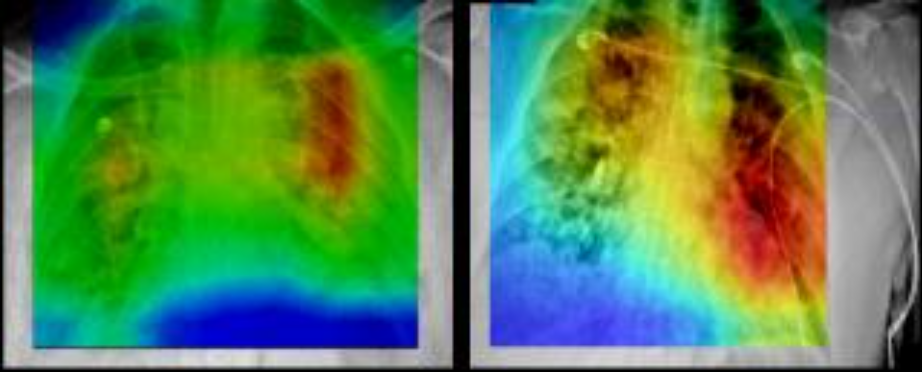
This collection of cases was acquired at Stony Brook University from patients who tested positive for COVID-19. The collection includes images from different modalities and organ sites (chest radiographs, chest CTs, brain MRIs, etc.). Radiology imaging data is extremely important in COVID-19 from both a diagnostic and a monitoring perspective, given the crucial nature of COVID-19 pulmonary disease and its rapid...
This is a sample collection of synthetic 3D Digital Reference Objects (DROs) intended for standardization of quantitative imaging feature extraction pipelines. We have developed a software toolkit for the creation of DROs with customizable size, shape, intensity, texture, and margin sharpness values. Using user-supplied input parameters, these objects are defined mathematically...
PROSTATEx has been superseded by PI-CAI:
The ProstateX dataset (both training and testing cases) have been included in the PI-CAI Public Training and Development dataset. As such, ProstateX as a benchmark has been deprecated and is superseded by the PI-CAI challenge. PI-CAI is an all-new grand challenge, with over 10,000 carefully-curated prostate MRI exams to validate modern AI algorithms and estimate...
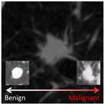
As part of the 2015 SPIE Medical Imaging Conference, SPIE – with the support of American Association of Physicists in Medicine (AAPM) and the National Cancer Institute (NCI) – will conduct a “Grand Challenge” on quantitative image analysis methods for the diagnostic classification of malignant and benign lung nodules. The LUNGx Challenge will provide a unique opportunity for participants to compare their algorithms...
Read More
Microscopic images were captured from bone marrow aspirate slides of patients diagnosed with B-lineage Acute Lymphoid Leukemia (B-ALL) and Multiple Myeloma (MM) as per the standard guidelines. Slides were stained using Jenner-Giemsa stain. Images were captured at 1000x magnification using Nikon Eclipse-200 microscope equipped with a digital camera. Images were captured in raw BMP format with a size of 2560x1920 pixels....

This collection contains longitudinal DCE MRI studies of 64 patients undergoing neoadjuvant chemotherapy (NACT) for invasive breast cancer.
Patient population
This pilot study to investigate the use of serial DCE MRI examinations during neoadjuvant chemotherapy for invasive breast cancer recruited 68 patients with stage II or III locally advanced breast cancer enrolled between...
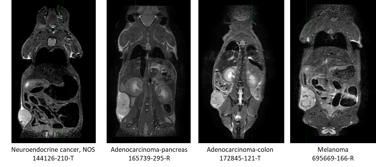
This collection contains serial non-contrast non-gated T2w MRI of 18 patient derived xenograft cancer models. 175 mice were imaged at multiple time points (514 total studies) for researchers to develop algorithms using neural networks, and classification techniques to improve tissue characterization (morphological changes) for the improvement in patient care through advances in precision medicine.
Characterization...
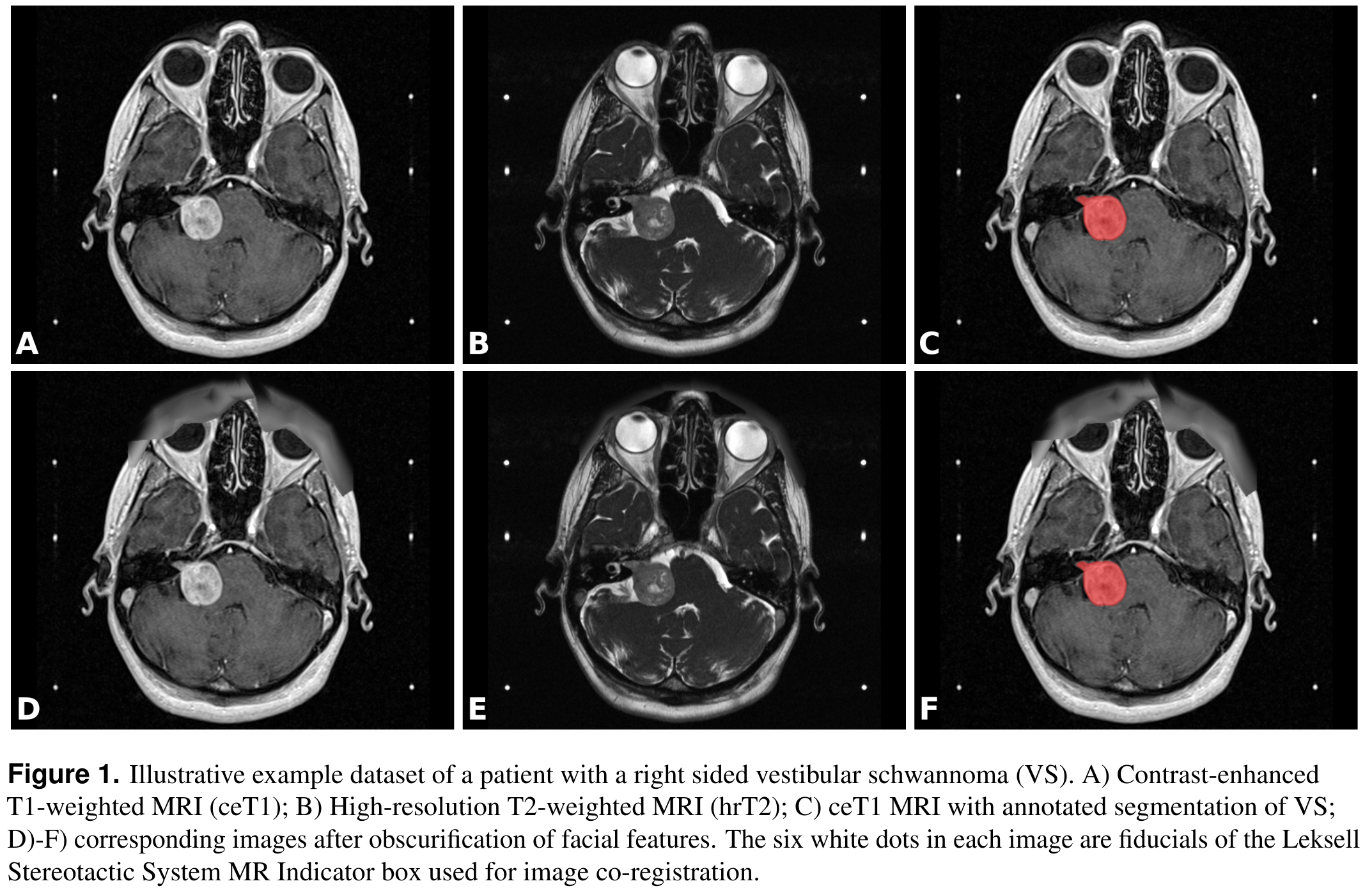
This collection contains a labeled dataset of MRI images collected on 242 consecutive patients with vestibular schwannoma (VS) undergoing Gamma Knife stereotactic radiosurgery (GK SRS). The structural images included contrast-enhanced T1-weighted (ceT1) images and high-resolution T2-weighted (hrT2) images. Each imaging dataset is accompanied by the patient’s radiation therapy (RT) dataset including the RTDose, RTStructures...