
VESTIBULAR-SCHWANNOMA-MC-RC

By
DOI: 10.7937/HRZH-2N82 | Image Collection

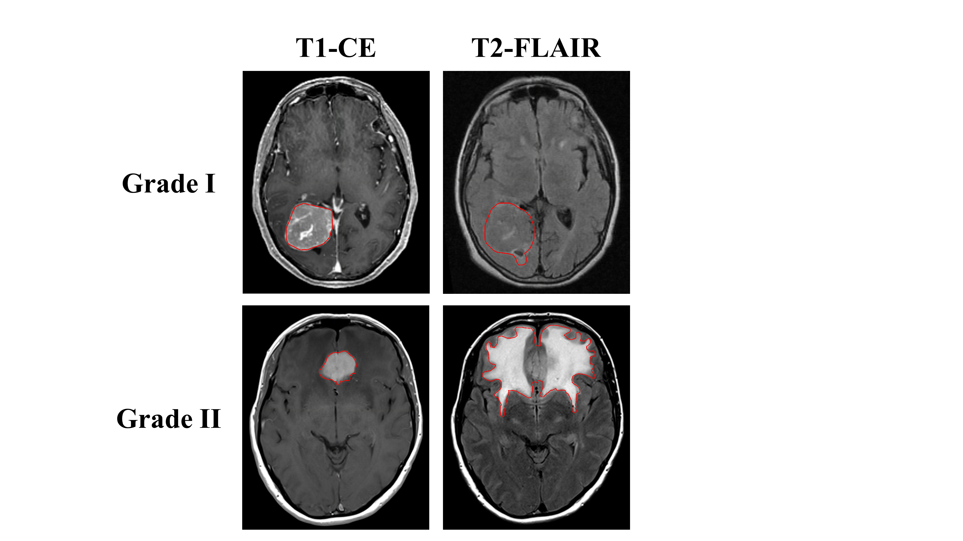
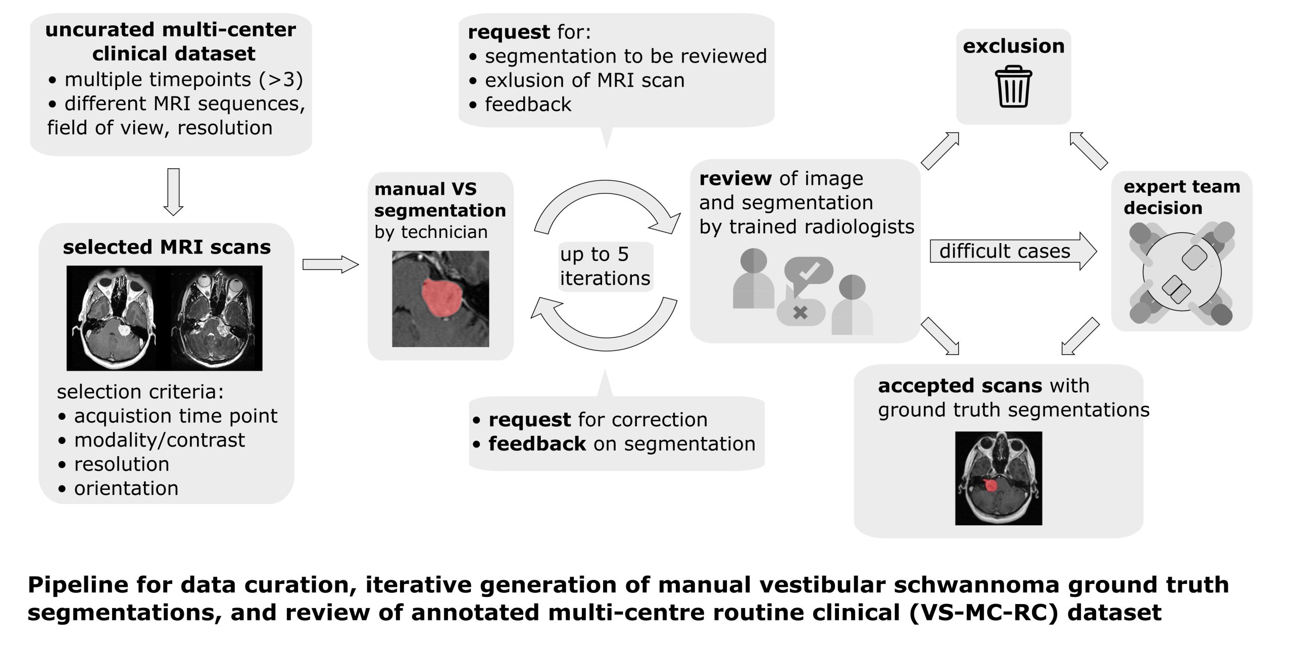
The Vestibular-Schwannoma-MC-RC (VS-MC-RC) dataset contains longitudinal MRI scans of 160 patients with a single sporadic Vestibular Schwannoma (VS) from 10 medical sites in the United Kingdom. For all patients either a T1-weighted (T1w) or T2-weighted (T2w) scan, or both, are available. The dataset provides binary segmentations of the VS for all scan sessions. The dataset comprises up to three time points for each...

Read More

MENINGIOMA-SEG-CLASS

By
DOI: 10.7937/0TKV-1A36 | Image Collection

The study included 96 consecutive treatment naïve patients with intracranial meningiomas treated with surgical resection from 2010 to 2019. All patients had pre-operative T1, T1-CE, and T2-FLAIR MR images with subsequent subtotal or gross total resection of pathologically confirmed grade I or grade II meningiomas. A neuropathology team reviewed histopathology, including two subspecialty trained neuropathologists and...

Read More

RIDER-PHANTOM-PET-CT

By
DOI: 10.7937/K9/TCIA.2015.8WG2KN4W | Image Collection

The RIDER PHANTOM PET-CT collection consists of repeat measurement PET/CT phantom scan collections carried out under the aegis of the Society of Nuclear Medicine (SNM) to discern the uniformity of clinical imaging instrumentation at various sites. They were obtained in cooperation with SNM as a resource for increased quantitative understanding of machine acquisition, analytic reproducibility and image processing.

The...

Read More

RIDER-PHANTOM-MRI

By
DOI: 10.7937/k9/tcia.2015.mi4qddhu | Image Collection

The RIDER Phantom MRI data set contains repeat phantom studies. The phantom used for all data acquisitions was a version of the EuroSpin II Test Object 5 as distributed by Diagnostic Sonar, Ltd (Livingston, West Lothian, Scotland). The phantom was comprised of 18 25-mm doped gel filled tubes and 1 20-mm tube containing 0.25 mM GdDTPA.

Scanners evaluated:

  • Scanner A – 1.5T GE 8-channel HD with BRM...

Read More

RIDER-NEURO-MRI

By
DOI: 10.7937/K9/TCIA.2015.VOSN3HN1 | Image Collection

RIDER Neuro MRI contains imaging data on 19 patients with recurrent glioblastoma who underwent repeat imaging sets.  These images were obtained approximately 2 days apart (with the exception of one patient, RIDER Neuro MRI-1086100996, whose images were obtained one day apart).   

DCE‐MRI:  All 19 patients had repeat dynamic contrast‐enhanced MRI (DCE‐MRI) datasets on the same...

Read More

RIDER-LUNG-PET-CT

By
DOI: 10.7937/k9/tcia.2015.ofip7tvm | Image Collection

 The RIDER Lung PET-CT collection was shared to facilitate the RIDER PET/CT subgroup activities. The PET/CT subgroup was responsible for: (1) archiving de-identified DICOM serial PET/CT phantom and lung cancer patient data in a public database to provide a resource for the testing and development of algorithms and imaging tools used for assessing response to therapy, (2) conducting multiple serial imaging studies...

Read More