PROSTATEx-Seg-Zones | PROSTATEx Zone Segmentations
DOI: 10.7937/TCIA.NBB4-4655 | Data Citation Required | Analysis Result
| Location | Subjects | Size | Updated | |||
|---|---|---|---|---|---|---|
| Prostate | Prostate | 98 | Organ segmentations | 2020/11/25 |
Summary
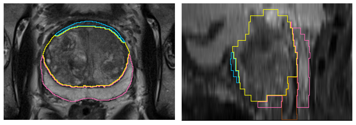
This collection contains prostate’s zonal segmentation for 98 cases randomly selected from the SPIE-AAPM-NCI PROSTATEx Challenges (PROSTATEx). The four-class segmentation encompasses the peripheral zone, transition zone, fibromuscular stroma and the distal prostatic urethra. As underlying images, we used transversal T2w scans. Segmentations were created by a medical student with experience in prostate segmentation and an expert urologist who instructed the student and double-checked the segmentations in the end. The DICOM representation of these segmentations were generated with dcmqi.
Data Access
Version 1: Updated 2020/11/25
| Title | Data Type | Format | Access Points | Subjects | License | |||
|---|---|---|---|---|---|---|---|---|
| Segmentations | SEG | DICOM | Download requires NBIA Data Retriever |
98 | 98 | 98 | 98 | CC BY 3.0 |
Collections Used In This Analysis Result
| Title | Data Type | Format | Access Points | Subjects | License | |||
|---|---|---|---|---|---|---|---|---|
| Corresponding Original MR Images from PROSTATEx | MR | DICOM | Requires NBIA Data Retriever |
98 | 98 | 98 | 2,020 | CC BY 3.0 |
Citations & Data Usage Policy
Data Citation Required: Users must abide by the TCIA Data Usage Policy and Restrictions. Attribution must include the following citation, including the Digital Object Identifier:
Data Citation |
|
|
Meyer, A., Schindele, D., von Reibnitz, D., Rak, M., Schostak, M., & Hansen, C. (2020). PROSTATEx Zone Segmentations [Data set]. The Cancer Imaging Archive. https://doi.org/10.7937/TCIA.NBB4-4655 |
Acknowledgements
This work has been funded by the EU and the federal state of Saxony-Anhalt, Germany under grant number ZS/2016/08/80388. We would like to thank the authors of the PROSTATEx challenge for providing this valuable dataset.
Related Publications
Publication Citation |
|
|
Meyer, A., Rakr, M., Schindele, D., Blaschke, S., Schostak, M., Fedorov, A., & Hansen, C. (2019, April). Towards Patient-Individual PI-Rads v2 Sector Map: Cnn for Automatic Segmentation of Prostatic Zones From T2-Weighted MRI. 2019 IEEE 16th International Symposium on Biomedical Imaging (ISBI 2019). 2019 IEEE 16th International Symposium on Biomedical Imaging (ISBI). https://doi.org/10.1109/isbi.2019.8759572 |
Research Community Publications
TCIA maintains a list of publications which leverage TCIA data. If you have a manuscript you’d like to add please contact the TCIA Helpdesk.