ISPY1-Tumor-SEG-Radiomics | Expert tumor annotations and radiomic features for the ISPY1/ACRIN 6657 trial data collection
DOI: 10.7937/TCIA.XC7A-QT20 | Data Citation Required | 758 Views | 3 Citations | Analysis Result
| Location | Subjects | Size | Updated | |||
|---|---|---|---|---|---|---|
| Breast Cancer | Breast | 163 | Tumor segmentations, radiomic features | 2022/06/01 |
Summary
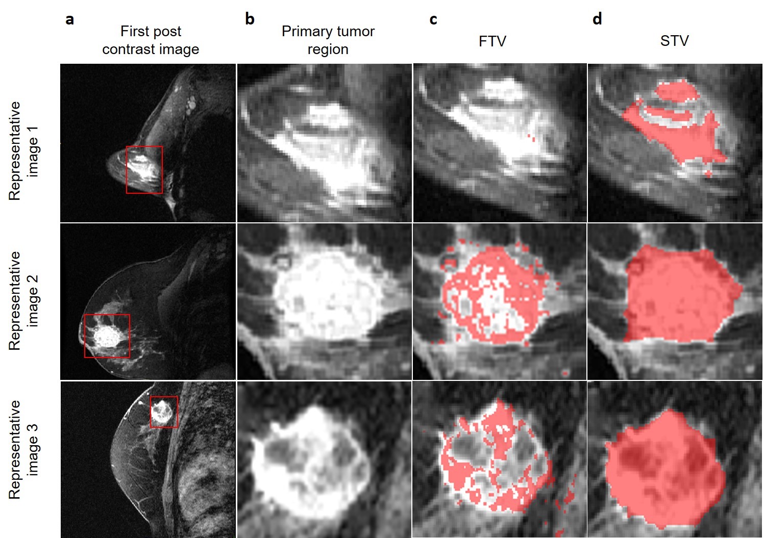
This dataset enhances the ISPY1 data collection, with uniformly curated data, tumor annotations, and quantitative imaging features. This dataset includes a) uniformly processed scans that are harmonized to match the intensity and spatial characteristics, facilitating immediate use in computational studies, b) computationally-generated and manually-revised expert annotations of tumor regions, as well as c) a comprehensive set of quantitative imaging (also known as radiomic) features corresponding to the tumor regions. The segmentations for the ISPY1/ACRIN 6657 dataset currently hosted on TCIA’s website describe a) the tumor volume of interest (VOI) and b) functional tumor volume (FTV). These currently provided segmentations do not include non-enhancing portions of the tumor volume, which represent a significant portion of the disease burden that needs to be studied to better understand and quantify the disease. The segmentations in these new analysis results are for the entire 3D primary lesion, including both the enhancing and the non-enhancing tumor regions, therefore defining the structural tumor volume (STV). These STV annotations were generated by manually delineating the primary lesion volume, after confirming the location of the primary lesion from the provided VOI and FTV. The STV annotations were reviewed and approved by a board-certified, fellowship-trained breast radiologist, and are statistically significantly different from FTV. We believe these STV annotations will allow analyses of the entire disease burden and analyses of tumor heterogeneity regarding contrast uptake, contributing to further expanding our mechanistic understanding of the disease potentially leading to improved patient management.
Data Access
Version 1: Updated 2022/06/01
| Title | Data Type | Format | Access Points | Subjects | License | Metadata | |||
|---|---|---|---|---|---|---|---|---|---|
| Images and Segmentations | MR, Segmentation | NIFTI | Download requires IBM-Aspera-Connect plugin |
163 | 1,467 | CC BY 3.0 | — | ||
| Radiomics Features | Radiomic Feature | XLSX | CC BY 3.0 | — | |||||
| README File | Other | TXT | CC BY 3.0 | — | |||||
| CaPTk radiomic feature parameter | Radiomic Feature | CSV | CC BY 3.0 | — |
Collections Used In This Analysis Result
| Title | Data Type | Format | Access Points | Subjects | License | Metadata | |||
|---|---|---|---|---|---|---|---|---|---|
| Corresponding Original MR Cases from ISPY1 | MR | DICOM | Requires NBIA Data Retriever |
163 | 629 | 4,898 | 298,304 | CC BY 3.0 | View |
Citations & Data Usage Policy
Data Citation Required: Users must abide by the TCIA Data Usage Policy and Restrictions. Attribution must include the following citation, including the Digital Object Identifier:
Data Citation |
|
|
Chitalia, R., Pati, S., Bhalerao, M., Thakur, S., Jahani, N., Belenky, J. V., McDonald, E.S., Gibbs, J., Newitt, D., Hylton, N., Kontos, D., & Bakas, S. (2021). Expert tumor annotations and radiomic features for the ISPY1/ACRIN 6657 trial data collection [Data set]. The Cancer Imaging Archive. https://doi.org/10.7937/TCIA.XC7A-QT20 |
Acknowledgements
Research reported in this publication was partly supported by the National Cancer Institute (NCI) of the National Institutes of Health (NIH), under award numbers U01CA242871 and U24CA189523, U01CA151235, R01CA197000, and R01CA132870.
Related Publications
Publications by the Dataset Authors
The authors recommended the following as the best source of additional information about this dataset:
Publication Citation |
|
|
Chitalia, R., Pati, S., Bhalerao, M., Thakur, S. P., Jahani, N., Belenky, V., McDonald, E. S., Gibbs, J., Newitt, D. C., Hylton, N. M., Kontos, D., & Bakas, S. (2022). Expert tumor annotations and radiomics for locally advanced breast cancer in DCE-MRI for ACRIN 6657/I-SPY1. In Scientific Data (Vol. 9, Issue 1). Springer Science and Business Media LLC. https://doi.org/10.1038/s41597-022-01555-4 |
Research Community Publications
TCIA maintains a list of publications that leveraged this dataset. If you have a manuscript you’d like to add please contact TCIA’s Helpdesk.