Breast cancer (BC) is the second most commonly diagnosed cancer in the U.S. with more than 250,000 new cases of invasive breast cancers reported in 2017. The majority of women with locally advanced and a subset of patients with operable breast cancer will undergo systemic therapy prior to their surgery (neoadjuvant therapy/ NAT) to reduce the size of tumor(s) and possibly further undergo breast conserving surgery....
The Munich AML Morphology Dataset contains 18,365 expert-labeled single-cell images taken from peripheral blood smears of 100 patients diagnosed with Acute Myeloid Leukemia at Munich University Hospital between 2014 and 2017, as well as 100 patients without signs of hematological malignancy. Image acquisition was done using a M8 digital microscope / scanner (Precipoint GmbH, Freising, Germany) at 100-fold optical magnification...
This collection contains FDG-PET/CT and anatomical MR (T1-weighted, T2-weighted with fat-suppression) imaging data from 51 patients with histologically proven soft-tissue sarcomas (STSs) of the extremities. All patients had pre-treatment FDG-PET/CT and MRI scans between November 2004 and November 2011. (Note: date in the TCIA images have been changed in the interest of de-identification; the same change was applied...
...
...
The dataset contains a collection of over 170,000 de-identified, expert-annotated cells from the bone marrow smears of 945 patients stained using the May-Grünwald-Giemsa/Pappenheim stain. The diagnosis distribution in the cohort included a variety of hematological diseases reflective of the sample entry of a large laboratory specialized in leukemia diagnostics. Image acquisition was performed using a brightfield microscope...
This collection consists of Computed Tomography (CT) images of the mediastinum and abdomen in which lymph node positions are marked by radiologists at the National Institutes of Health, Clinical Center. Radiologists at the Imaging Biomarkers and Computer-Aided Diagnosis Laboratory labeled a total...
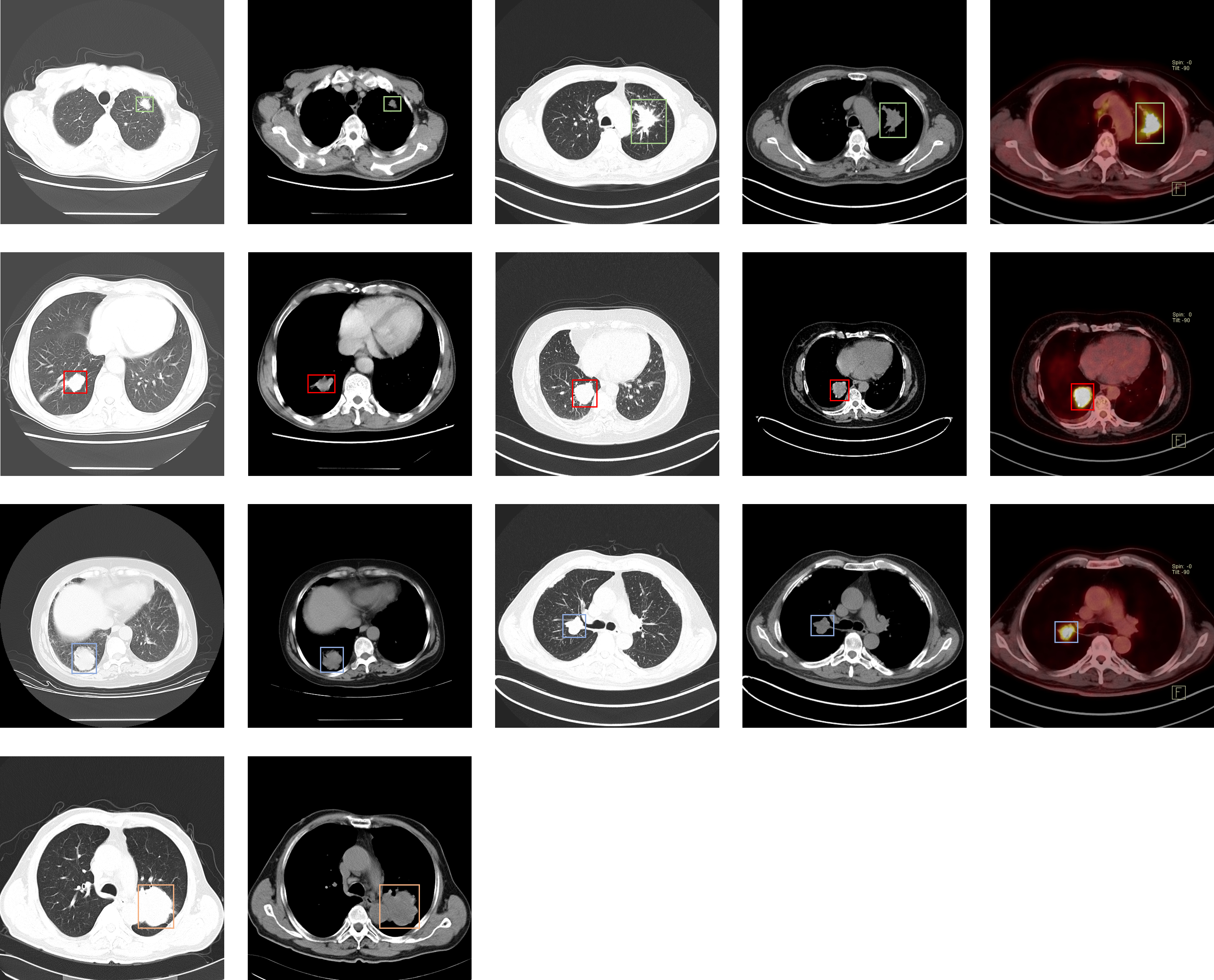
This dataset consists of CT and PET-CT DICOM images of lung cancer subjects with XML Annotation files that indicate tumor location with bounding boxes. The images were retrospectively acquired from patients with suspicion of lung cancer, and who underwent standard-of-care lung biopsy and PET/CT. Subjects were grouped according to a tissue histopathological diagnosis. Patients with Names/IDs containing the letter...
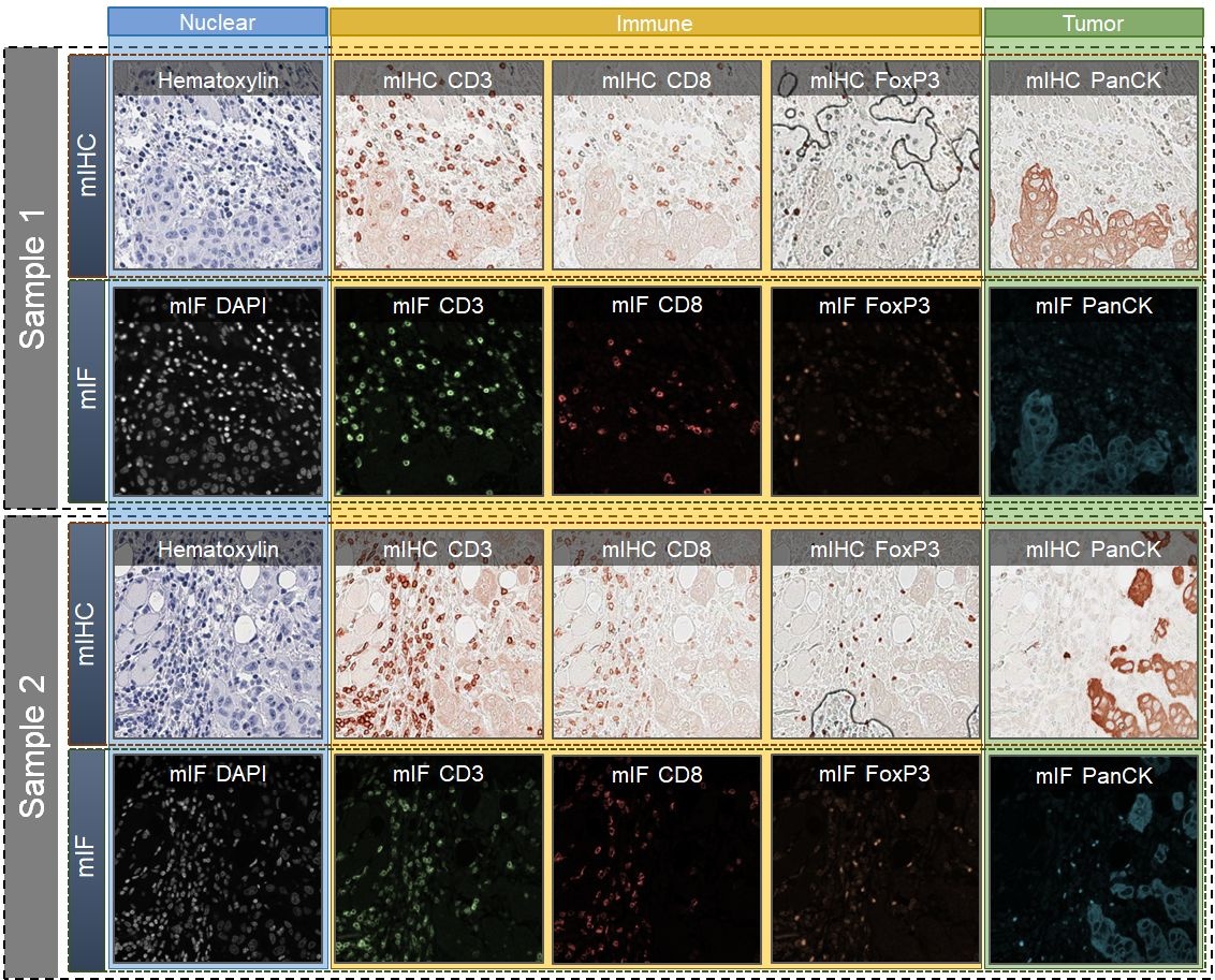
We introduce a new AI-ready computational pathology dataset containing restained and co-registered digitized images from eight head-and-neck squamous cell carcinoma patients. Specifically, the same tumor sections were stained with the expensive multiplex immunofluorescence (mIF) assay first and then restained with cheaper multiplex immunohistochemistry (mIHC). This is a first public dataset that demonstrates...