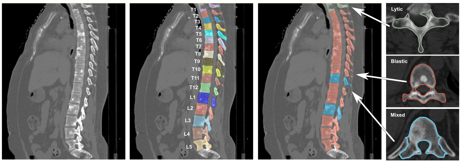
Spine-Mets-CT-SEG | Spine metastatic bone cancer: pre and post radiotherapy CT
DOI: 10.7937/kh36-ds04 | Data Citation Required | 6k Views | Image Collection
| Location | Species | Subjects | Data Types | Cancer Types | Size | Status | Updated | |
|---|---|---|---|---|---|---|---|---|
| Bone | Human | 55 | Demographic, Follow-Up, Classification, CT, SEG | Metastatic disease, Bladder Cancer, Breast Cancer, Colon Cancer, Kidney Cancer, Lung Cancer, Prostate Cancer, Soft-tissue Sarcoma, Skin Cancer | Clinical | Public, Complete | 2024/09/25 |
Summary
We provide an annotated imaging dataset of cancerous CT spines to help develop artificial intelligence frameworks for automatic vertebrae segmentation and classification. This collection contains a dataset of 55 CT scans collected on patients with a large range of primary cancers and corresponding bone metastatic lesions obtained for patients with metastatic spine disease. The subjects of the study planned for radiotherapy were simulated at the Radiation Oncology Department, Brigham and Women's Hospital, Boston, MA, using 1) Siemens SOMATOM Confidence (Siemens Healthcare GmbH, Erlangen, Germany) and 2) GE Lightspeed (General Electric Medical System, Waukesha, WI) CT scanner. Simulation scan parameters are detailed in Table 1. Table 1. CT image acquisition parameters. Radiotherapy CT scanner Siemens SOMATOM Confidence GE Lightspeed Protocol parameters SBRT All Others SBRT All Others kVp 120 120 120 120 mA Variable Variable 300 300 FOV A*, B** A*, B** A*, B** A*, B** Slice Thickness 0.5mm 1.5mm 1.25mm 1.25mm In-Plane Pixel Size (mm) 0.31x0.31 0.31x0.31 0.31x0.31 0.31x0.31 Gantry rotation 1s 1s 1s 1s Gating None None None None Breath Hold None None None None *A: 16cm field of view, **B: Skin-to-Skin field of view The dataset includes: See Usage Note in Detailed Description section for more detail.
Data Access
Version 1: Updated 2024/09/25
| Title | Data Type | Format | Access Points | Subjects | License | Metadata | |||
|---|---|---|---|---|---|---|---|---|---|
| Images | CT, SEG | DICOM | Download requires NBIA Data Retriever |
55 | 55 | 110 | 35,582 | CC BY 4.0 | View |
| Spine Lesion Classifications and Demographics | Demographic, Follow-Up, Classification | XLSX | 55 | CC BY 4.0 | — |
Additional Resources for this Dataset
The NCI Cancer Research Data Commons (CRDC) provides access to additional data and a cloud-based data science infrastructure that connects data sets with analytics tools to allow users to share, integrate, analyze, and visualize cancer research data.
- Imaging Data Commons (IDC) (Imaging Data)
Citations & Data Usage Policy
Data Citation Required: Users must abide by the TCIA Data Usage Policy and Restrictions. Attribution must include the following citation, including the Digital Object Identifier:
Data Citation |
|
|
Pieper, S., Haouchine, N., Hackney, D.B., Wells, W.M. Sanhinova, M., Balboni, T., Spektor, A., Huynh, M., Tanguturi, S., Kim, E., Guenette, J.P., Kozono, D.E., Czajkowski, B., Caplan, S., Doyle, P., Kang, H., Alkalay, R.N. (2024) Spine metastatic bone cancer: pre and post radiotherapy CT (Spine-Mets-CT-SEG) [Dataset] (Version 1). The Cancer Imaging Archive. https://doi.org/10.7937/kh36-ds04 |
Detailed Description
Usage note:
We recommend 3D Slicer’s DICOM viewer (https://www.slicer.org), to load and view the DICOM images. The CT images can be viewed without additional extensions. The segmentations can be viewed using the QuantitativeReporting extensions (https://qiicr.gitbook.io/quantitativereporting-guide/)
If needed, you can use the script tcia_dcm2nifit.py (https://github.com/rouge1616/Spine-Mets-CT-SEG/) to convert the full dataset to NIfTI file format.
Acknowledgements
This data set is supported by funding from
- NATIONAL INSTITUTE OF ARTHRITIS AND MUSCULOSKELETAL AND SKIN DISEASES , grant # 3R01AR075964-03S1
- NATIONAL INSTITUTE OF ARTHRITIS AND MUSCULOSKELETAL AND SKIN DISEASES, grant # R01AR075964
Harmonization of the components of this dataset, including into standard DICOM representation, was supported in part by the NCI Imaging Data Commons consortium. NCI Imaging Data Commons consortium is supported by the contract number 19X037Q from Leidos Biomedical Research under Task Order HHSN26100071 from NCI.
The SAIC Metadata Content Development Team identified existing or created new caDSR Common Data elements (CDEs) to describe the harmonized components of this dataset. The Metadata Content Development Team is supported by CBIIT under Task Order 140D0421F0008 from NCI.
Related Publications
Publications by the Dataset Authors
The authors recommended the following as the best source of additional information about this dataset:
Publication Citation |
|
|
Fourney, D. R., Frangou, E. M., et al. (2011). Spinal Instability Neoplastic Score: An Analysis of Reliability and Validity From the Spine Oncology Study Group. In Journal of Clinical Oncology (Vol. 29, Issue 22, pp. 3072–3077). American Society of Clinical Oncology (ASCO). https://doi.org/10.1200/jco.2010.34.3897 |
.
Research Community Publications
TCIA maintains a list of publications that leveraged this dataset. If you have a manuscript you’d like to add please contact TCIA’s Helpdesk.
