MiMM_SBILab | MiMM_SBILab Dataset: Microscopic Images of Multiple Myeloma
DOI: 10.7937/tcia.2019.pnn6aypl | Data Citation Required | Image Collection
| Location | Species | Subjects | Data Types | Cancer Types | Size | Status | Updated | |
|---|---|---|---|---|---|---|---|---|
| Bone | Human | 5 | Histopathology | Multiple Myeloma | Image Analyses | Public, Complete | 2019/03/25 |
Summary
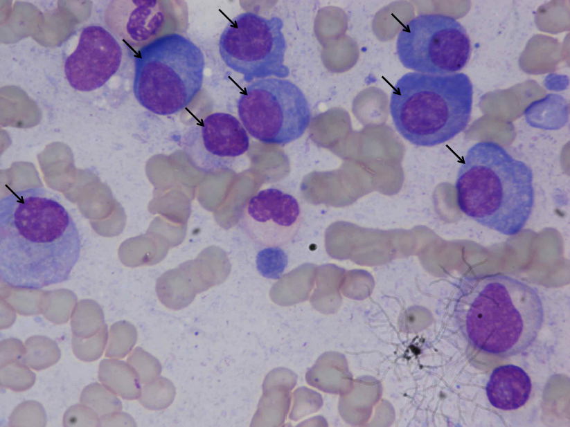
Microscopic images were captured from bone marrow aspirate slides of patients diagnosed with multiple myeloma as per the standard guidelines. Slides were stained using Jenner- Giemsa stain. Images were captured at 1000x magnification using Nikon Eclipse-200 microscope equipped with a digital camera. Images were captured in raw BMP format with a size of 2560x1920 pixels. In all, this dataset consists of 85 images. All these 85 images were stain normalized using our in-house methodology before being used for segmentation. These stain normalized images have been provided as the annotated dataset with plasma cells marked in all image slides contained in a presentation for the ready reference of readers. This collection has also been uploaded to the Harvard Blood Cancer Dataverse website. Please refer to DOI 10.7910/DVN/XCX7ST for more information.Additional Notes
Data Access
Version 1: Updated 2019/03/25
| Title | Data Type | Format | Access Points | Subjects | License | |||
|---|---|---|---|---|---|---|---|---|
| Slide Images | Histopathology | BMP | Download requires IBM-Aspera-Connect plugin |
5 | 5 | 85 | CC BY 3.0 | |
| Annotated plasma cell images | CC BY 3.0 |
Citations & Data Usage Policy
Data Citation Required: Users must abide by the TCIA Data Usage Policy and Restrictions. Attribution must include the following citation, including the Digital Object Identifier:
Data Citation |
|
|
Gupta, R., & Gupta, A. (2019). MiMM_SBILab Dataset: Microscopic Images of Multiple Myeloma [Data set]. The Cancer Imaging Archive. https://doi.org/10.7937/tcia.2019.pnn6aypl |
Related Publications
Publication Citation |
|
|
Gupta, A., Duggal, R., Gehlot, S., Gupta, R., Mangal, A., Kumar, L., Thakkar, N., & Satpathy, D. (2020). GCTI-SN: Geometry-inspired chemical and tissue invariant stain normalization of microscopic medical images. Medical Image Analysis, 65, 101788. https://doi.org/10.1016/j.media.2020.101788 |
Publication Citation |
|
|
Gupta, A., Mallick, P., Sharma, O., Gupta, R., & Duggal, R. (2018). PCSeg: Color model driven probabilistic multiphase level set based tool for plasma cell segmentation in multiple myeloma. PLOS ONE, 13(12), e0207908. https://doi.org/10.1371/journal.pone.0207908 |
Research Community Publications
The following publications are recommended by the data submitters that may be useful to researchers utilizing this collection:
- Ritu Gupta, Pramit Mallick, Rahul Duggal, Anubha Gupta, and Ojaswa Sharma, “Stain Color Normalization and Segmentation of Plasma Cells in Microscopic Images as a Prelude to Development of Computer Assisted Automated Disease Diagnostic Tool in Multiple Myeloma,” 16th International Myeloma Workshop (IMW), India, March 2017 https://doi.org/10.1016/j.clml.2017.03.178
TCIA maintains a list of publications which leverage TCIA data. If you have a manuscript you’d like to add please contact TCIA’s Helpdesk.