Healthy-Total-Body-CTs | Low-Dose CT Images of Healthy Cohort
DOI: 10.7937/NC7Z-4F76 | Data Citation Required | 6.3k Views | 1 Citations | Image Collection
| Location | Species | Subjects | Data Types | Cancer Types | Size | Status | Updated | |
|---|---|---|---|---|---|---|---|---|
| Whole body | Human | 30 | CT, Segmentation, Demographic | Non-Cancer | Clinical, Image Analyses | Limited, Complete | 2024/09/27 |
Summary
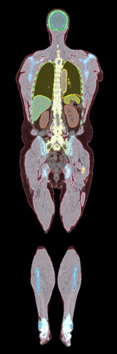
This data set includes low-dose whole body CT images and tissue segmentations of thirty healthy adult research participants who underwent PET/CT imaging on the uEXPLORER total-body PET/CT system at UC Davis. Participants included in this study were healthy adults, 18 years of age or older, who were able to provide informed written consent. The participants' age, sex, weight, height, and body mass index are also provided. Fifteen participants underwent PET/CT imaging at three timepoints during a 3-hour period (0 minutes, 90 minutes, and 180 minutes) after PET radiotracer injection, while the remaining 15 participants were imaged at six timepoints during a 12-hour period (additionally at 360 minutes, 540 minutes, and 720 minutes). The imaging timepoint is indicated in the Series Description DICOM tag, with a value of either 'dyn', '90min', '3hr', '6hr', '9hr', or '12hr', corresponding to the delay after PET tracer injection. CT images were acquired immediately before PET image acquisition. Currently, only CT images are included in the data set from either three or six timepoints. The tissue segmentations include 37 tissues consisting of 13 abdominal organs, 20 different bones, subcutaneous and visceral fat, skeletal and psoas muscle. Segmentations were automatically generated at the 90 minute timepoint for each participant using MOOSE, an AI segmentation tool for whole body data. The segmentations are provided in NIFTI format and may need to be re-oriented to correctly match the CT image data in DICOM format. The uEXPLORER CT scanner is an 80-row, 160 slice CT scanner typically used for anatomical imaging and attenuation correction for PET/CT. The CT scan obtained at 90 minutes was performed with 140 kVp and an average of 50 mAs for all subjects. At all other time-points (0 minutes, 180 minutes, etc.) the CT scan was obtained with 140 kVp and an average of 5 mAs. CT images were reconstructed into a 512x512x828 image matrix with 0.9766x0.9766x2.344 mm3 voxel size. A key is provided along with the segmentations download in the Data Access table which details the organ values.
Data Access
Some data in this collection contains images that could potentially be used to reconstruct a human face. To safeguard the privacy of participants, users must sign and submit a TCIA Restricted License Agreement to help@cancerimagingarchive.net before accessing the data.
Version 2: Updated 2024/09/27
- All scans for a single patient took place on the same day, dates were modified to reflect this where previously they did not.
- A single date was selected for all date data 2002/06/19. The original dates in the dataset had been modified; there was not a correlation to the actual acquisition or creation dates in the imaging metadata.
- Time in any date/time metadata was maintained at original value.
- All data for a single patient is now in the same study. Originally, each timepoint for a single patient was assigned to a study; there were multiple studies for a single patient.
- Patient age and sex were added to metadata where missing.
- Clinical data spreadsheet corrected. Patient IDs were incorrect, updated to match metadata. Corrected height column title from centimeters (cm) to meters (m).
| Title | Data Type | Format | Access Points | Subjects | License | Metadata | |||
|---|---|---|---|---|---|---|---|---|---|
| Images | CT | DICOM | Download requires NBIA Data Retriever |
30 | 30 | 135 | 111,778 | TCIA Restricted | View |
| Segmentations & Segmentation Organ Values spreadsheet | Segmentation | NIFTI, XLSX, and ZIP | 30 | 30 | CC BY 4.0 | — | |||
| Clinical Data | Demographic | XLSX | CC BY 4.0 | — |
Citations & Data Usage Policy
Data Citation Required: Users must abide by the TCIA Data Usage Policy and Restrictions. Attribution must include the following citation, including the Digital Object Identifier:
Data Citation |
|
|
Selfridge, A. R., Spencer, B., Shiyam Sundar, L. K., Abdelhafez, Y., Nardo, L., Cherry, S. R., & Badawi, R. D. (2023). Low-Dose CT Images of Healthy Cohort (Healthy-Total-Body-CTs) (Version 2) [Dataset]. The Cancer Imaging Archive. https://doi.org/10.7937/NC7Z-4F76 |
Acknowledgements
We would like to acknowledge the individuals and institutions that have provided data for this collection:
Funding for this work was provided by NIH grant R01 CA206187, which is supported by NCI, NIBIB and the Office of the Director, and by R01 CA249422.
Related Publications
Publications by the Dataset Authors
The authors recommended the following as the best source of additional information about this dataset:
Publication Citation |
|
|
Sundar, L. K. S., Yu, J., Muzik, O., Kulterer, O. C., Fueger, B., Kifjak, D., Nakuz, T., Shin, H. M., Sima, A. K., Kitzmantl, D., Badawi, R. D., Nardo, L., Cherry, S. R., Spencer, B. A., Hacker, M., & Beyer, T. (2022). Fully Automated, Semantic Segmentation of Whole-Body18F-FDG PET/CT Images Based on Data-Centric Artificial Intelligence. In Journal of Nuclear Medicine (Vol. 63, Issue 12, pp. 1941–1948). Society of Nuclear Medicine. https://doi.org/10.2967/jnumed.122.264063 |
No other publications were recommended by dataset authors.
Research Community Publications
TCIA maintains a list of publications that leveraged this dataset. If you have a manuscript you’d like to add please contact TCIA’s Helpdesk.
Previous Versions
Version 1: Updated 2023/09/21
| Title | Data Type | Format | Access Points | License | Metadata | ||||
|---|---|---|---|---|---|---|---|---|---|
| Images | CT | DICOM | Download requires NBIA Data Retriever |
30 | 30 | 135 | 111,778 | TCIA Restricted | — |
| Segmentations & Segmentation Organ Values spreadsheet | Segmentation | NIFTI, XLSX, and ZIP | 30 | 30 | CC BY 4.0 | — | |||
| Clinical Data | Demographic | XLSX | CC BY 4.0 | — |