HE-vs-MPM | Multimodal imaging of ductal carcinoma in situ with microinvasion
DOI: 10.7937/3fyc-ac78 | Data Citation Required | Image Collection
| Location | Species | Subjects | Data Types | Cancer Types | Size | Status | Updated |
|---|---|---|---|---|---|---|---|
| Breast | Human | 12 | Histopathology | Breast Cancer | Public, Complete | 2023/12/08 |
Summary
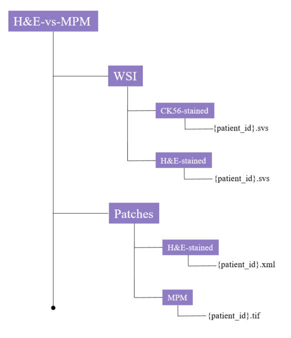
Ductal carcinoma in situ with microinvasion (DCISM) is a challenging subtype of breast cancer with controversial invasiveness and prognosis. Accurate diagnosis of DCISM from ductal carcinoma in situ (DCIS) is crucial for optimal treatment and improved clinical outcomes. This dataset provides histopathology images and paired CK5/6 immunohistochemical staining images from patients with DCISM, as well as multiphoton microscopy images of suspicious regions. It offers multi-modal imaging data from various perspectives for analysis and diagnosis of microinvasive breast cancer by other researchers in the field. The dataset contains data from 12 breast cancer patients, including 10 cases of ductal carcinoma in situ with microinvasion (DCISM), 1 case of ductal carcinoma in situ (DCIS), and 1 case of invasive breast cancer. The magnification of the glass slide images is 40x. The pathology slide scanner used was created by the Sunny Optical Technology (group) Co., Ltd., and the pixel aspect ratio of the images is 1. The dataset also includes multiphoton microscopy imaging of suspicious microinvasion areas. The multiphoton imaging system was manufactured by Zeiss, and it also has a pixel aspect ratio of 1. Our database was specifically collected for the use of imaging methods in diagnosing DICSM. The suffixes in each case number indicate the patient's condition - "DCISM" for ductal carcinoma in situ with microinvasion, "DCIS" for ductal carcinoma in situ, and "IDC" for invasive ductal carcinoma. Apart from these labels, we have not collected any additional clinical information for these cases.
Data Access
Version 1: Updated 2023/12/08
| Title | Data Type | Format | Access Points | Subjects | License | |||
|---|---|---|---|---|---|---|---|---|
| Tissue Slide Images | Histopathology | SVS, TIFF, and XML | Download requires IBM-Aspera-Connect plugin |
12 | 116 | CC BY 4.0 |
Citations & Data Usage Policy
Data Citation Required: Users must abide by the TCIA Data Usage Policy and Restrictions. Attribution must include the following citation, including the Digital Object Identifier:
Data Citation |
|
|
Han, X., Liu, Y., Chen, J., & Kang, D. (2023). Multimodal imaging of ductal carcinoma in situ with microinvasion (HE-vs-MPM) (Version 1) [Data set]. The Cancer Imaging Archive. https://doi.org/10.7937/3FYC-AC78 |
Acknowledgements
- This work was supported by the National Natural Science Foundation of China (Grant No. 82171991), Natural Science Foundation of Fujian Province (Nos. 2020J01154, 2020J011008, 2020J01839, 2022J01170), Joint Funds for the Innovation of Science and Technology of Fujian Province (2019Y9101), the special Funds of the Central Government Guiding Local Science and Technology Development (No. 2020L3008), and Fujian Major Scientific and Technological Special Project for “Social Development” (No. 2020YZ016002).
Related Publications
Publication Citation |
|
|
Han, X., Liu, Y., Zhang, S., Li, L., Zheng, L., Qiu, L., Chen, J., Zhan, Z., Wang, S., Ma, J., Kang, D., & Chen, J. (2024). Improving the diagnosis of ductal carcinoma in situ with microinvasion without immunohistochemistry: An innovative method with H&E‐stained and multiphoton microscopy images. In International Journal of Cancer (Vol. 154, Issue 10, pp. 1802–1813). Wiley. https://doi.org/10.1002/ijc.34855 |
Research Community Publications
TCIA maintains a list of publications which leverage TCIA data. If you have a manuscript you’d like to add please contact TCIA’s Helpdesk.