Adrenal-ACC-Ki67-Seg | Voxel-level segmentation of pathologically-proven Adrenocortical carcinoma with Ki-67 expression
DOI: 10.7937/1FPG-VM46 | Data Citation Required | Image Collection
| Location | Species | Subjects | Data Types | Cancer Types | Size | Status | Updated | |
|---|---|---|---|---|---|---|---|---|
| Adrenal | Human | 53 | SEG, CT, Demographic, Diagnosis, Follow-Up, Measurement | Adrenocortical Carcinoma | Clinical, Image Analyses | Public, Complete | 2023/05/15 |
Summary
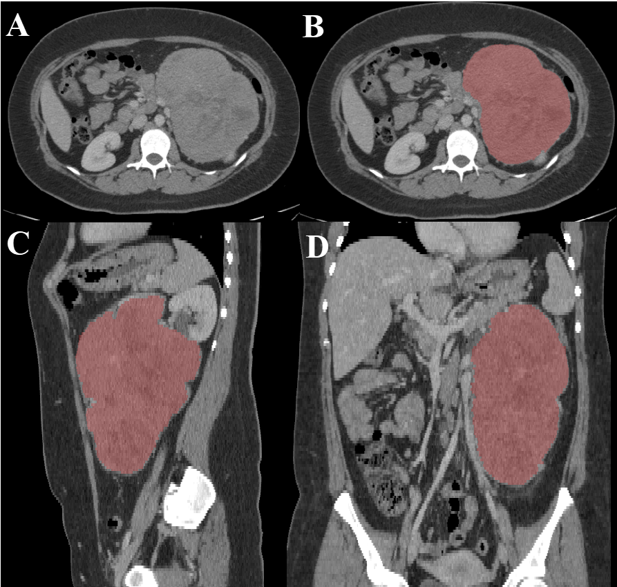
Adrenocortical carcinoma (ACC) is a rare tumor of the adrenal cortex with a reported annual incidence of one case per million population. ACC is a highly aggressive, highly fatal tumor with 5-year overall survival rates ranging from 14% to 44%. Diagnosis of ACC is primarily based on histopathological parameters from resected tumors, which include Ki-67 expression status. The Ki-67 index is one of the most important established prognostic markers for local recurrence of ACC. Radiomic feature extraction showed a significant association between radiomic signature and Ki-67 expression status in ACC. This retrospectively acquired data includes contrast enhanced CT imaging studies of 53 confirmed ACC patients between 2006 to 2018 with available clinical and pathological data, including Ki-67 index. Semi-automatic segmentation of the adrenal tumor was created using AMIRA, then manually refined by an experienced radiologist. Voxel level segmentation of the adrenal lesion are included as well. The segmentations of each contrast-enhanced CT were done for the purpose of radiomic features extraction. The participants in this dataset fulfilled these inclusion criteria: Data from patients whose Ki-67 was quantified in biopsied tissue samples rather than from resected whole tumor, were excluded from this study. This exclusion was based on previous studies concluding that Ki-67 quantification should be based on tissue samples collected from the whole tumour. There was no publicly-available library for adrenal lesions prior to this dataset. It can serve as a training set for machine learning algorithms for various purposes including segmentation and classification of adrenal tumors. We used the radiomic features extracted to predict the Ki-67 index (through regression) without the need of surgical intervention as described in this paper.
Data Access
Version 1: Updated 2023/05/15
| Title | Data Type | Format | Access Points | Subjects | License | |||
|---|---|---|---|---|---|---|---|---|
| Images and Segmentations | SEG, CT | DICOM | Download requires NBIA Data Retriever |
53 | 65 | 177 | 18,255 | CC BY 4.0 |
| Clinical data | Demographic, Diagnosis, Follow-Up, Measurement | XLSX | CC BY 4.0 |
Additional Resources for this Dataset
The NCI Cancer Research Data Commons (CRDC) provides access to additional data and a cloud-based data science infrastructure that connects data sets with analytics tools to allow users to share, integrate, analyze, and visualize cancer research data.
- Imaging Data Commons (IDC) (Imaging Data, Digitized Histopathology)
Citations & Data Usage Policy
Data Citation Required: Users must abide by the TCIA Data Usage Policy and Restrictions. Attribution must include the following citation, including the Digital Object Identifier:
Data Citation |
|
|
Moawad, A. W., Ahmed, A. A., ElMohr, M., Eltaher, M., Habra, M. A., Fisher, S., Perrier, N., Zhang, M., Fuentes, D., & Elsayes, K. (2023). Voxel-level segmentation of pathologically-proven Adrenocortical carcinoma with Ki-67 expression (Adrenal-ACC-Ki67-Seg) [Data set]. The Cancer Imaging Archive. https://doi.org/10.7937/1FPG-VM46 |
Acknowledgements
- The University of Texas MD Anderson Cancer Center, departments of Surgical Oncology, Endocrinology, Pathology, Imaging Physics, and Diagnostic Radiology.
- The authors would like to thank the Scientific Publication department at the University of Texas MD Anderson Cancer Center for their contribution to this dataset and articles.
Related Publications
Publications by the Dataset Authors
The authors recommended this paper as the best source of additional information about this dataset:
Ahmed, A. A., Elmohr, M. M., Fuentes, D., Habra, M. A., Fisher, S. B., Perrier, N. D., Zhang, M., & Elsayes, K. M. (2020). Radiomic mapping model for prediction of Ki-67 expression in adrenocortical carcinoma. In Clinical Radiology (Vol. 75, Issue 6, p. 479.e17-479.e22). Elsevier BV. https://doi.org/10.1016/j.crad.2020.01.012
No publications by dataset authors were found.
Research Community Publications
TCIA maintains a list of publications which leverage our data. If you have a manuscript you’d like to add please contact the TCIA Helpdesk.